File list
Jump to navigation
Jump to search
This special page shows all uploaded files.
| Date | Name | Thumbnail | Size | User | Description | Versions |
|---|---|---|---|---|---|---|
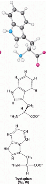
| 14:19, 29 November 2013 | Tryptophan.png (file) |  |
82 KB | 130024257 (talk | contribs) | Structure of Tryptophan | 1 |
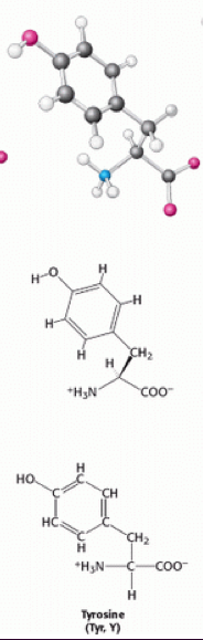
| 14:19, 29 November 2013 | Tyrosine.png (file) |  |
82 KB | 130024257 (talk | contribs) | Structure of Tyrosine | 1 |
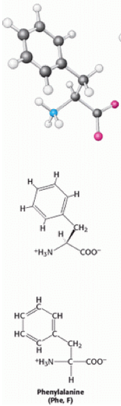
| 14:17, 29 November 2013 | Phenylalanine.png (file) |  |
81 KB | 130024257 (talk | contribs) | Structure of Phenylalanine | 1 |
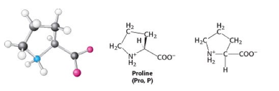
| 14:17, 29 November 2013 | Proline.png (file) |  |
34 KB | 130024257 (talk | contribs) | Structure of Proline | 1 |
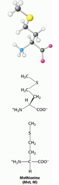
| 14:16, 29 November 2013 | Methionine.png (file) |  |
66 KB | 130024257 (talk | contribs) | Structure of Methionine | 1 |
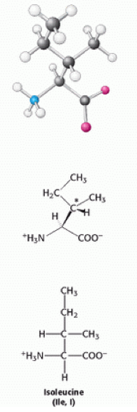
| 14:15, 29 November 2013 | Isoleucine.png (file) |  |
68 KB | 130024257 (talk | contribs) | Strucure of Isoleucine | 1 |
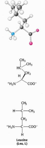
| 14:14, 29 November 2013 | Leucine.png (file) |  |
64 KB | 130024257 (talk | contribs) | Structure of Leucine | 1 |
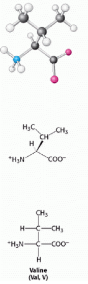
| 14:14, 29 November 2013 | Valine.png (file) |  |
60 KB | 130024257 (talk | contribs) | Strucutre of Valine | 1 |
| 14:11, 29 November 2013 | Alanine.png (file) |  |
29 KB | 130024257 (talk | contribs) | Structure of Alanine | 1 |
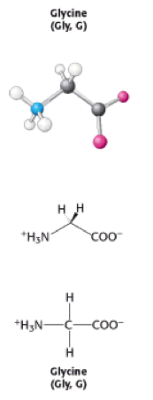
| 14:00, 29 November 2013 | Glycine.png (file) |  |
27 KB | 130024257 (talk | contribs) | Structure of glycine | 1 |
| 18:36, 28 November 2013 | Actin filament.jpg (file) |  |
36 KB | 130186739 (talk | contribs) | Reverted to version as of 18:27, 28 November 2013 | 10 |
| 16:48, 28 November 2013 | Griffith's experiment of transformation.jpg (file) | 35 KB | 130098993 (talk | contribs) | Griffith's Experiment of Bacterial Transformation | 1 | |
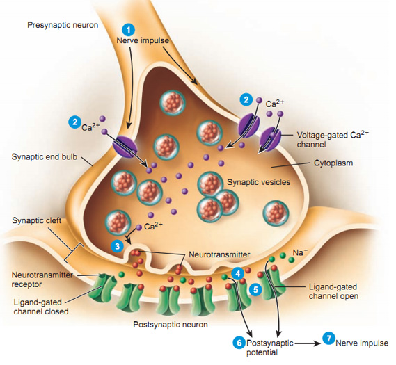
| 17:38, 27 November 2013 | Synapse.jpg (file) |  |
102 KB | 130071860 (talk | contribs) | 2 | |
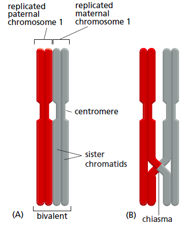
| 15:36, 27 November 2013 | Screen Shot 2013-11-27 at 15.23.19.png (file) |  |
37 KB | 130059075 (talk | contribs) | On the left: A bivalent structure normally joined by synaptonemal complex. On the right: Crossover has happened between non-sister chromatids. A chiasma is identified at the point which connects the homologs. This diagram was reproduced from page 1274 of | 1 |
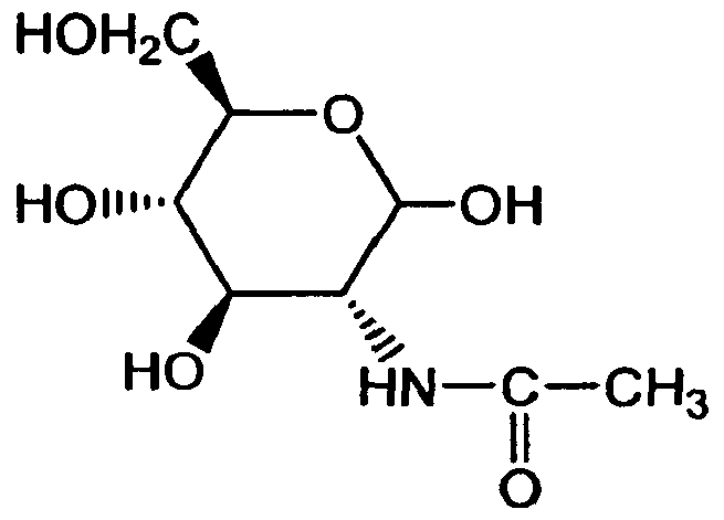
| 15:15, 27 November 2013 | Imgf000019 0001.png (file) |  |
4 KB | 120030848 (talk | contribs) | N-acetylglucosamine structure | 1 |
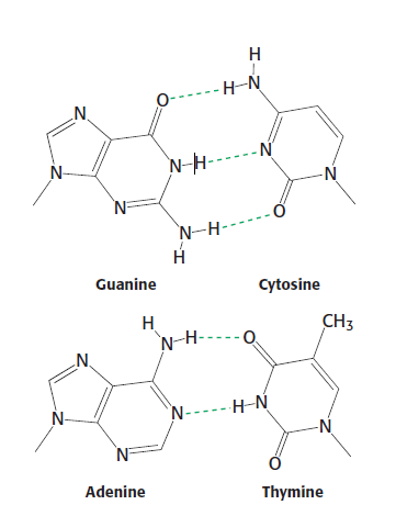
| 10:14, 25 November 2013 | Watson-Crick Base Pairs.PNG (file) |  |
18 KB | 140001705 (talk | contribs) | Structures of the base pairs proposed by Watson and Crick. | 1 |
| 23:59, 14 November 2013 | Phagocytosis.jpg (file) |  |
70 KB | 130366423 (talk | contribs) | Image showing the process of Phagocytosis. | 1 |
| 12:16, 14 November 2013 | Karyotype-clg-10980.jpg (file) |  |
21 KB | 120066065 (talk | contribs) | Human female karyotype, consisting of 44 autosomes plus 2 allosomes designated XX. | 1 |
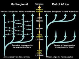
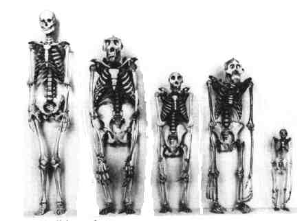
| 19:55, 12 November 2013 | Multiregional and out of africa model .jpg (file) |  |
11 KB | 130052180 (talk | contribs) | This image shows a summary of the Multiregional model and the Africa replacement model. | 2 |
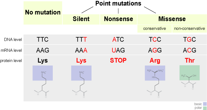
| 22:46, 8 November 2013 | Point mutations-en.png (file) |  |
32 KB | 130043834 (talk | contribs) | 2 | |
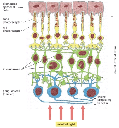
| 16:51, 23 October 2013 | Retina.jpg (file) |  |
128 KB | 110026626 (talk | contribs) | The neural structure of the retina. Notice the layered organisation of the cells and the fact that those cells that transmit the signals to the brain lie closest to the light stimulus. The stimulation of the photoreceptors (either rods or cones) by light | 1 |
| 16:40, 20 October 2013 | Screen Shot 2013-10-20 at 17.30.44.png (file) |  |
50 KB | 110075255 (talk | contribs) | 1 | |
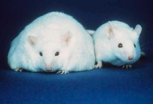
| 19:21, 14 October 2013 | Fatmouse-public-300x203.jpg (file) |  |
12 KB | 110188267 (talk | contribs) | Transgenic mouse with the MC4R gene knocked out against a normal mouse. | 1 |
| 00:28, 26 September 2013 | Eggsmilk.jpg (file) |  |
1.18 MB | Nnjm2 (talk | contribs) | Eggs and milk | 1 |
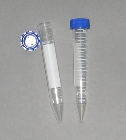
| 07:54, 12 April 2013 | 15 ml tube.png (file) |  |
177 KB | Nnjm2 (talk | contribs) | Test uplaod | 1 |
| 16:46, 30 November 2012 | SSU front and LSU front side by side white backgroundSMALL.jpg (file) |  |
313 KB | 120550645 (talk | contribs) | 30S and 50S rRNA | 1 |
| 16:45, 30 November 2012 | Mgsupdivision.jpg (file) |  |
92 KB | 120052781 (talk | contribs) | 1 | |
| 14:51, 30 November 2012 | B-D-Glucopyranose.jpg (file) |  |
11 KB | 110188267 (talk | contribs) | The chair and boat forms of b-D-glucopyranose | 1 |
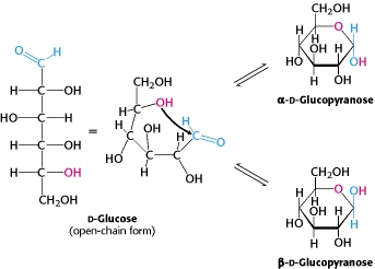
| 14:44, 30 November 2012 | D-glucose.jpg (file) |  |
28 KB | 110188267 (talk | contribs) | Open chain D glucose forming alpha and beta rings. | 1 |
| 12:36, 30 November 2012 | Divergence.png (file) |  |
72 KB | 110073491 (talk | contribs) | 1 | |
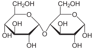
| 17:35, 29 November 2012 | Maltose.png (file) |  |
14 KB | 120223152 (talk | contribs) | The structure of maltose (Haworth projection) | 1 |
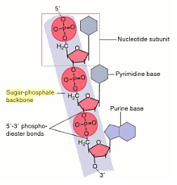
| 12:04, 29 November 2012 | Sugar Phosphate Backbone.png (file) |  |
13 KB | 120064935 (talk | contribs) | 1 | |
| 23:53, 22 November 2012 | Nuclear Receptors.jpg (file) | 67 KB | 120052437 (talk | contribs) | 1 | ||
| 17:08, 11 November 2012 | Scan1.jpg (file) |  |
174 KB | 120222948 (talk | contribs) | 2 | |
| 17:08, 11 November 2012 | Scan.jpg (file) |  |
196 KB | 120222948 (talk | contribs) | 1 | |
| 12:33, 27 October 2012 | 2012-08-28.jpg (file) |  |
473 KB | Nnjm2 (talk | contribs) | A bench | 1 |
| 12:18, 27 October 2012 | Aspartate5.gif (file) |  |
2 KB | Nnjm2 (talk | contribs) | 1 | |
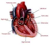
| 16:10, 2 December 2011 | Heart.jpg (file) |  |
5 KB | 110028697 (talk | contribs) | 1 | |
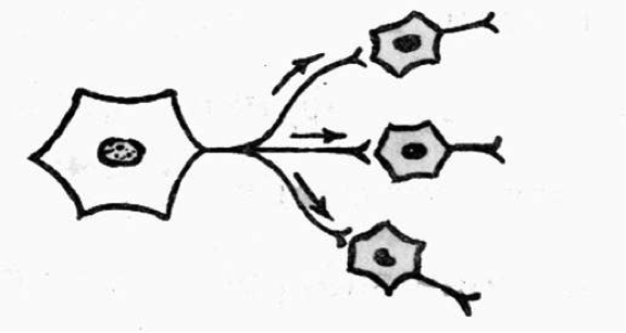
| 14:27, 2 December 2011 | Nervecell.gif (file) |  |
10 KB | 110335643 (talk | contribs) | labelled diagram of a nerve cell | 1 |
| 14:03, 2 December 2011 | Aminoacids.jpg (file) |  |
17 KB | 110028435 (talk | contribs) | 1 | |
| 12:01, 2 December 2011 | GeneticCodeTable.jpg (file) |  |
303 KB | 109292483 (talk | contribs) | Abstract from Faculty and Staff Personal http://facweb.northseattle.edu/estavney/Bio242/Lab/Graphics for Lectures and Lab/ | 1 |
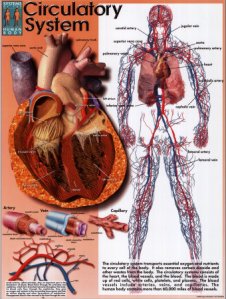
| 09:42, 2 December 2011 | Cardiovascularsystem.jpg (file) |  |
25 KB | 110028697 (talk | contribs) | The cardiovascular system | 2 |
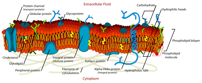
| 23:24, 1 December 2011 | Membrane Protein.png (file) |  |
199 KB | 110463094 (talk | contribs) | 1 | |
| 19:03, 1 December 2011 | Trisomy13.jpg (file) |  |
24 KB | 110156185 (talk | contribs) | Common symptoms of Trisomy 13 | 1 |
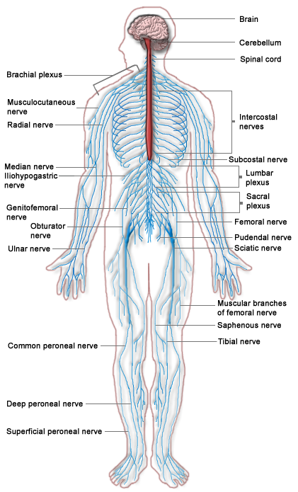
| 18:11, 1 December 2011 | Nervous system diagram.png (file) |  |
176 KB | 110029971 (talk | contribs) | 1 | |
| 16:54, 1 December 2011 | Skeleton.jpg (file) |  |
11 KB | 110104083 (talk | contribs) | 1 | |
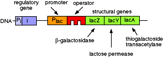
| 16:48, 1 December 2011 | Lac Operon.GIF (file) |  |
4 KB | 110102193 (talk | contribs) | The Lac-operon in E.coli | 1 |
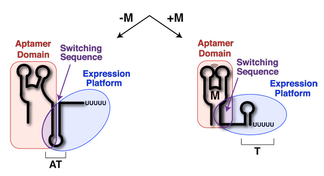
| 16:36, 1 December 2011 | Riboswitch.jpg (file) |  |
56 KB | 100412822 (talk | contribs) | A riboswitch showing translation is dependent on a lignad being bound to the riboswitch itself. | 1 |
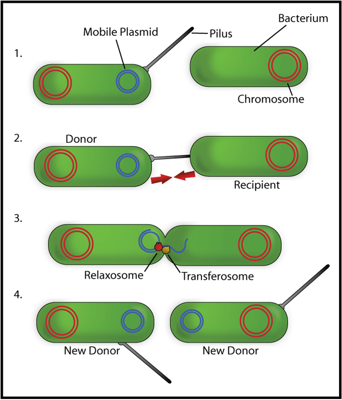
| 14:25, 1 December 2011 | Conjug.png (file) |  |
220 KB | 110163688 (talk | contribs) | 1 | |
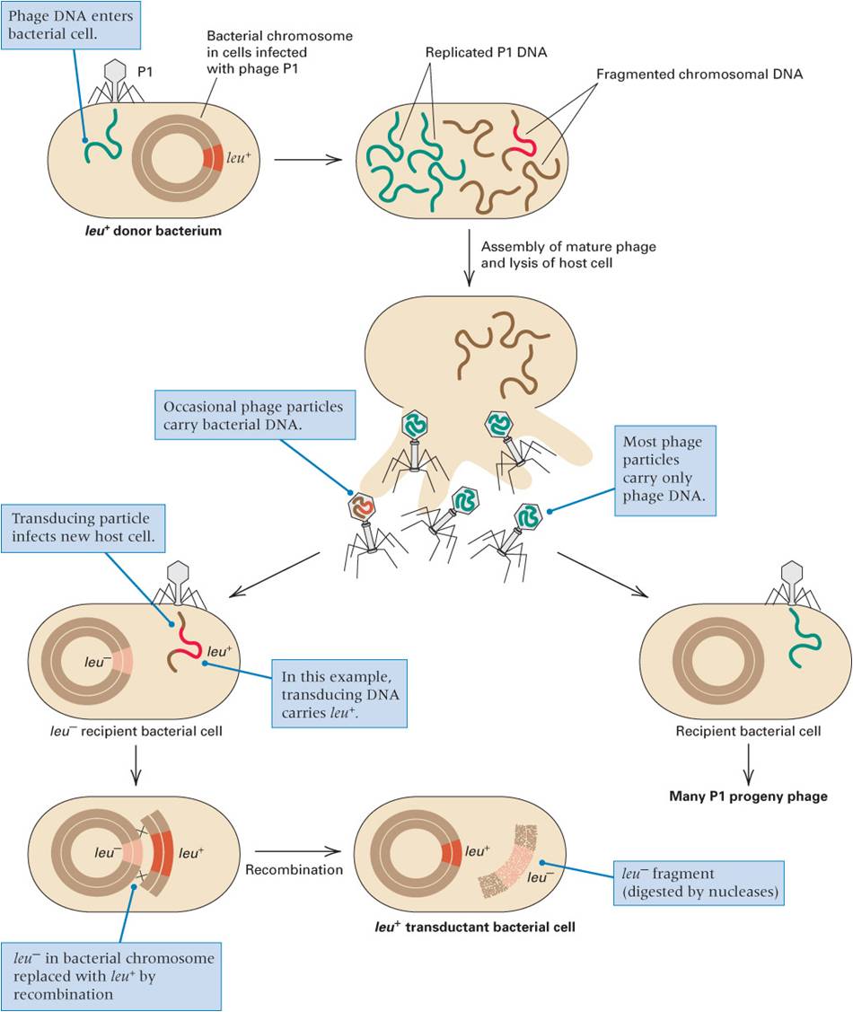
| 14:21, 1 December 2011 | Transduction.jpg (file) | 96 KB | 102997875 (talk | contribs) | 1 |