Smooth Muscle: Difference between revisions
No edit summary |
No edit summary |
||
| (3 intermediate revisions by 2 users not shown) | |||
| Line 1: | Line 1: | ||
[[ | [[Smooth muscle|Smooth muscle]] is the major involuntary contractile unit found within the Human body, with its main function being the regulation of content flow through hollow pathways. This muscle group is situated in varying, and vastly different areas if the body and include the internal diameter of small Arterioles and [[Blood vessels|blood]][[Blood vessels| vessels]]<span style="line-height: 1.5em;">, within the respiratory tract, stomach and intestines as well as the bladder and the eye<ref>Tortora, GJ., Derrickson, B. (2013) Essentials of Anatomy and Physiology Ninth Edition. Singapore: John Wiley & Sons, INC</ref>. Therefore, along with their major role of content regulation through hollow organs, smooth muscle cells, although the least specialised are also able to carry out a number of other roles</span><ref>Mackenna, BR., Callander, R. (1998) Illustrated Physiology Sixth Edition. Edinburgh: Churchill Livingstone pg 20</ref><span style="line-height: 1.5em;">. These include the focusing of the eye and the erecting of body hairs in response to temperature changes. </span> | ||
The muscle itself can be controlled by both electronic signals ([[Action potential|action potentials]]) and chemical signals ([[Hormones| | Regarding their major role, smooth muscle is able to carry out its functi<span style="line-height: 1.5em;">on by alternating between varied degrees of contraction and relaxation to control the dimensions of the organ. This results in an increase or decrease of resistance to flow of contents.</span> | ||
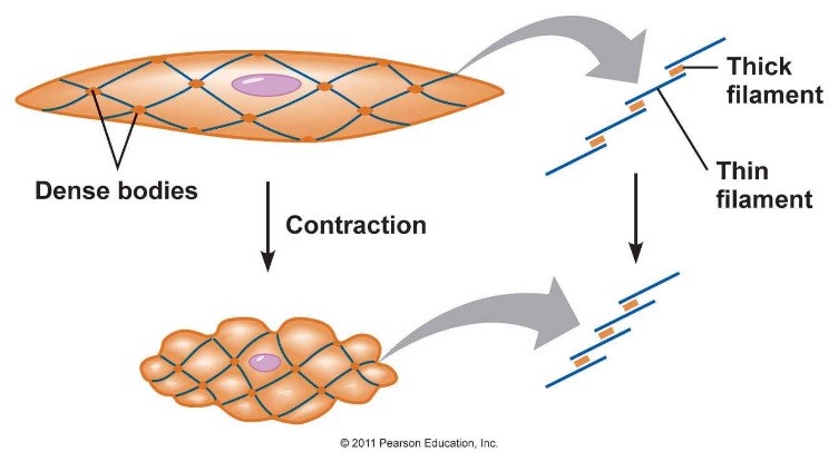
[[Image:Smooth_muscle_1.jpg|The contraction of a smooth muscle cell]]<br> | |||
'''STRUCTURE''' | |||
Smooth muscle is composed of many contractile muscle cells, with a single centrally-located nucleus. These cells contain [[Actin filaments|actin]] and [[Myosin|myosin]] filaments arranged in bundles as opposed to bands, meaning they differ from skeletal muscles by the fact that they have no striations or sarcomeres. Although the basic unit of contraction in these two types of muscle fibre (smooth muscle and [[Skeletal Muscle|skeletal muscle]]) are similar, smooth muscles l<span style="line-height: 1.5em;">ack Z lines, and are instead fixed in position by structres known as dense bodies(see above), The contraction of smooth muscles tends to be slower, but more prolonged than that of the skeletal muscles.</span> | |||
The muscle itself can be controlled by both electronic signals ([[Action potential|action potentials]]) and chemical signals <span style="line-height: 1.5em;">(</span>[[Hormones|hormo]][[Hormones|nes]]<span style="line-height: 1.5em;">) and, as such, may be divided into two sub-types. These are Single unit Smooth muscle - composed of electronically-coupled cells, where triggering contraction in one cell will induce contraction in all linked cells; and Multiunit Smooth muscle - composed of non-linked cells, so contraction can be triggered in one cell without causing stimulation in its neighbouring cells.</span><ref>Watras J.. (2008). Muscles. In: Koeppen B., Stanton B. Berne and Levy Physiology. Philadelphia: Mosby. p231-255.</ref><span style="line-height: 1.5em;"> </span> | |||
Smooth muscle activity patterns can vary, with some muscles able to carry out regular, rhythmic, 'wave-like' contraction and others constantly active (such as some sphincters)<ref>Medical physiology 2nd edition-walter F. Boron, Emile L. Boulpaep page253-262</ref>.<br> | Smooth muscle activity patterns can vary, with some muscles able to carry out regular, rhythmic, 'wave-like' contraction and others constantly active (such as some sphincters)<ref>Medical physiology 2nd edition-walter F. Boron, Emile L. Boulpaep page253-262</ref>.<br> | ||
Latest revision as of 19:39, 27 November 2014
Smooth muscle is the major involuntary contractile unit found within the Human body, with its main function being the regulation of content flow through hollow pathways. This muscle group is situated in varying, and vastly different areas if the body and include the internal diameter of small Arterioles and blood vessels, within the respiratory tract, stomach and intestines as well as the bladder and the eye[1]. Therefore, along with their major role of content regulation through hollow organs, smooth muscle cells, although the least specialised are also able to carry out a number of other roles[2]. These include the focusing of the eye and the erecting of body hairs in response to temperature changes.
Regarding their major role, smooth muscle is able to carry out its function by alternating between varied degrees of contraction and relaxation to control the dimensions of the organ. This results in an increase or decrease of resistance to flow of contents.
STRUCTURE
Smooth muscle is composed of many contractile muscle cells, with a single centrally-located nucleus. These cells contain actin and myosin filaments arranged in bundles as opposed to bands, meaning they differ from skeletal muscles by the fact that they have no striations or sarcomeres. Although the basic unit of contraction in these two types of muscle fibre (smooth muscle and skeletal muscle) are similar, smooth muscles lack Z lines, and are instead fixed in position by structres known as dense bodies(see above), The contraction of smooth muscles tends to be slower, but more prolonged than that of the skeletal muscles.
The muscle itself can be controlled by both electronic signals (action potentials) and chemical signals (hormones) and, as such, may be divided into two sub-types. These are Single unit Smooth muscle - composed of electronically-coupled cells, where triggering contraction in one cell will induce contraction in all linked cells; and Multiunit Smooth muscle - composed of non-linked cells, so contraction can be triggered in one cell without causing stimulation in its neighbouring cells.[3]
Smooth muscle activity patterns can vary, with some muscles able to carry out regular, rhythmic, 'wave-like' contraction and others constantly active (such as some sphincters)[4].
References
- ↑ Tortora, GJ., Derrickson, B. (2013) Essentials of Anatomy and Physiology Ninth Edition. Singapore: John Wiley & Sons, INC
- ↑ Mackenna, BR., Callander, R. (1998) Illustrated Physiology Sixth Edition. Edinburgh: Churchill Livingstone pg 20
- ↑ Watras J.. (2008). Muscles. In: Koeppen B., Stanton B. Berne and Levy Physiology. Philadelphia: Mosby. p231-255.
- ↑ Medical physiology 2nd edition-walter F. Boron, Emile L. Boulpaep page253-262
