File list
Jump to navigation
Jump to search
This special page shows all uploaded files.
| Date | Name | Thumbnail | Size | User | Description | Versions |
|---|---|---|---|---|---|---|
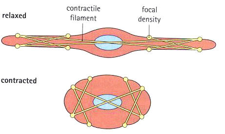
| 11:12, 30 November 2011 | Smooth muscle contraction.jpg (file) |  |
21 KB | 110029960 (talk | contribs) | Diagram showing smooth muscle contraction | 2 |
| 19:45, 29 November 2011 | Amyloid plaques.jpg (file) |  |
41 KB | 109292737 (talk | contribs) | 1 | |
| 19:43, 29 November 2011 | Alzhiemer's brain pix.jpg (file) |  |
9 KB | 109292737 (talk | contribs) | 1 | |
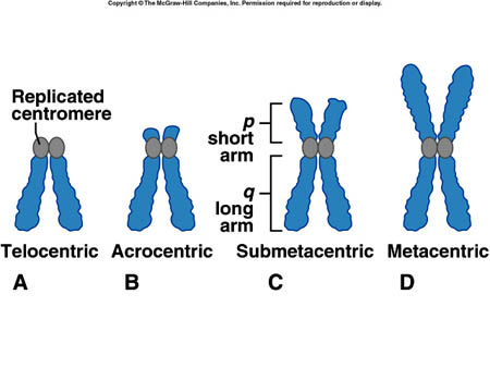
| 14:28, 29 November 2011 | Sister chromatids.jpg (file) |  |
57 KB | 109236410 (talk | contribs) | 1 | |
| 14:26, 29 November 2011 | Tp11 02.jpg (file) |  |
24 KB | 109236410 (talk | contribs) | 1 | |
| 12:21, 29 November 2011 | Mitochondrian.PNG (file) |  |
932 KB | 110443753 (talk | contribs) | A cross-section of a mitochondrion under an electron microscope | 1 |
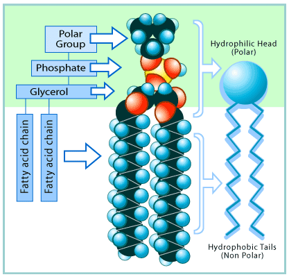
| 23:15, 28 November 2011 | Phospholipid.gif (file) |  |
33 KB | 110169509 (talk | contribs) | The structure of a phospholipid | 1 |
| 17:49, 27 November 2011 | Nucleosome 2.gif (file) |  |
64 KB | 100134658 (talk | contribs) | 1 | |
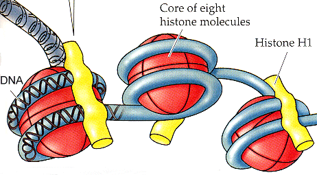
| 17:46, 27 November 2011 | Nucleosome.gif (file) |  |
64 KB | 100134658 (talk | contribs) | 1 | |
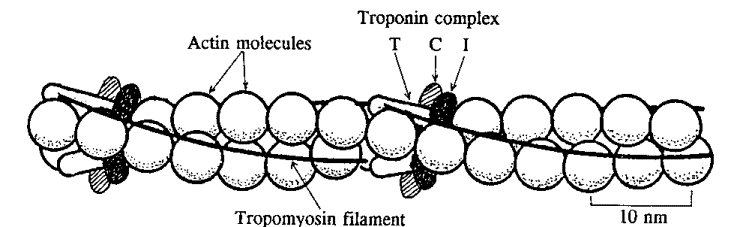
| 12:28, 27 November 2011 | Tropomyosin.gif (file) |  |
18 KB | 109276379 (talk | contribs) | Accessory proteins troponin and tropomyosin on the actin filament | 3 |
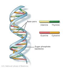
| 19:50, 26 November 2011 | BASE PAIRINGS.png (file) |  |
48 KB | 109292737 (talk | contribs) | 1 | |
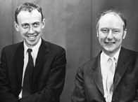
| 19:47, 26 November 2011 | Watson n crick.jpg (file) |  |
7 KB | 109292737 (talk | contribs) | James Watson anf Francis Crick | 1 |
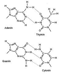
| 18:37, 26 November 2011 | BASE PAIRS.png (file) |  |
25 KB | 109292737 (talk | contribs) | A-T and G-C Base pairings | 1 |
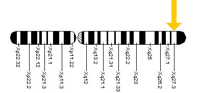
| 12:26, 24 November 2011 | X chromosome.jpg (file) |  |
12 KB | 110205748 (talk | contribs) | FMR-1 gene location | 1 |
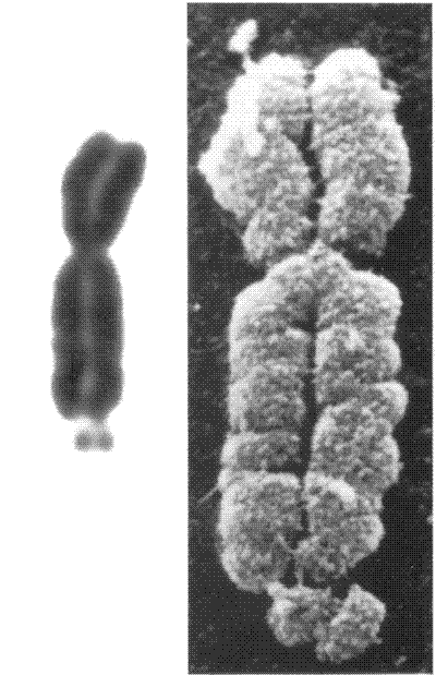
| 12:10, 24 November 2011 | Fragile X.gif (file) |  |
28 KB | 110205748 (talk | contribs) | A fragile X chromosome | 1 |
| 12:56, 21 November 2011 | Sliding filament theory.jpg (file) |  |
55 KB | 110054331 (talk | contribs) | The actin filaments slide past the myosin filaments toward the middle of the sarcomere (M line). The result is shortening of the sarcomere and muscle contraction, however there is no change in filament length. | 1 |
| 12:51, 21 November 2011 | Lrg-1348-skeletal muscle.jpg (file) |  |
28 KB | 110241436 (talk | contribs) | 1 | |
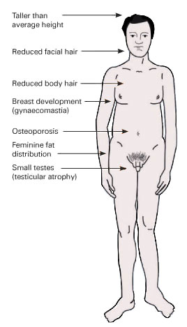
| 01:03, 20 November 2011 | KlinefeltersIMG.jpg (file) |  |
20 KB | 110091808 (talk | contribs) | Symptoms of Klinefelter syndrome | 1 |
| 23:43, 19 November 2011 | Piorb.gif (file) |  |
9 KB | 100234637 (talk | contribs) | 1 | |
| 23:42, 19 November 2011 | Tripple =.png (file) |  |
129 bytes | 100234637 (talk | contribs) | 1 | |
| 10:17, 19 November 2011 | Japan.gif (file) |  |
5 KB | 100802850 (talk | contribs) | ethnicity specific development of cancer in adults | 1 |
| 10:17, 19 November 2011 | Agr.gif (file) |  |
3 KB | 100802850 (talk | contribs) | age specific development of cancer in children | 1 |
| 10:16, 19 November 2011 | Age specific incidence.gif (file) |  |
4 KB | 100802850 (talk | contribs) | the age specific incidence of cancer in 30-80 year olds. | 1 |
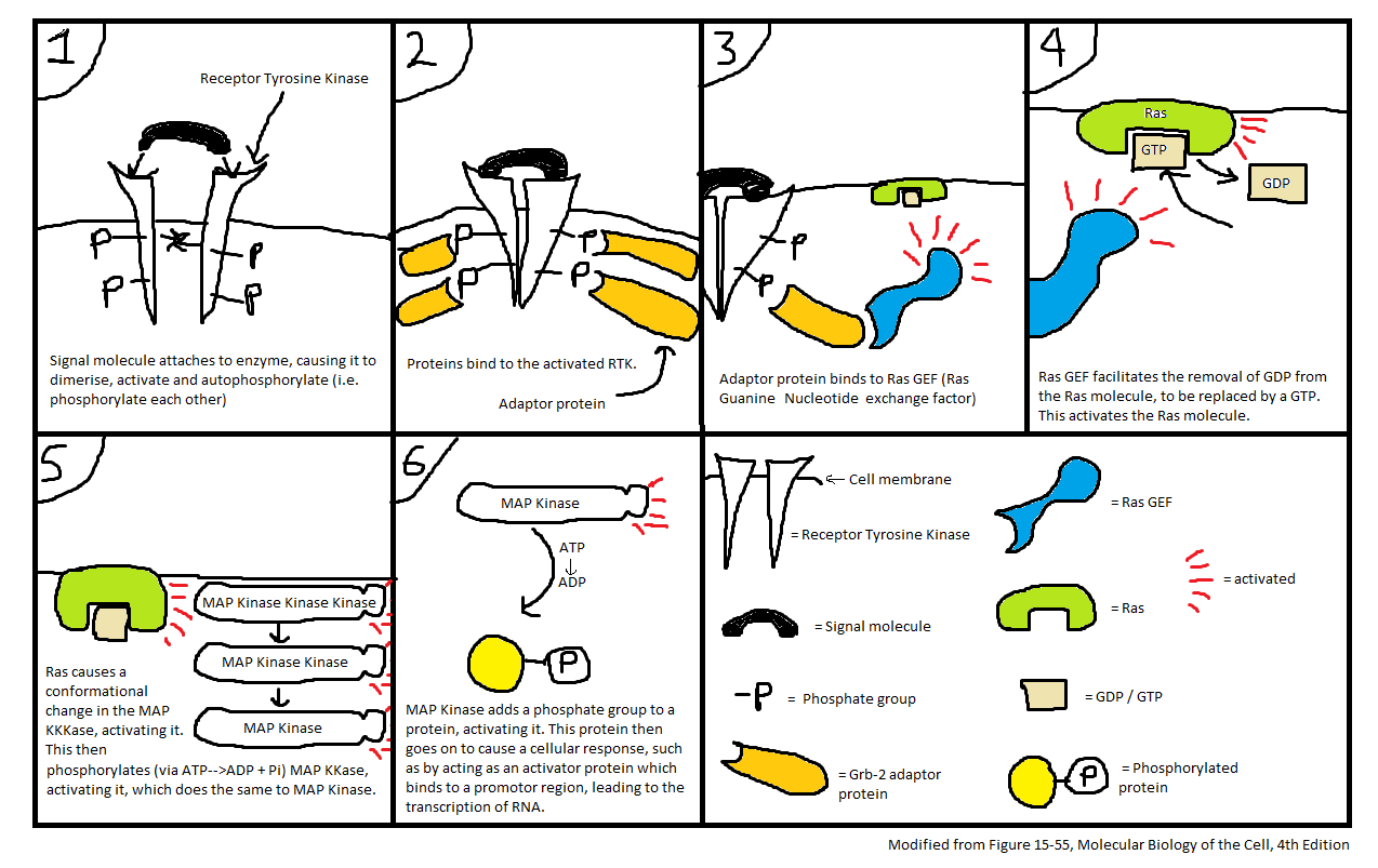
| 12:30, 17 November 2011 | Enzyme Linked Receptors (Ras).png (file) | .png) |
123 KB | 110028424 (talk | contribs) | A comic strip to show how the Ras signalling pathway takes place. | 1 |
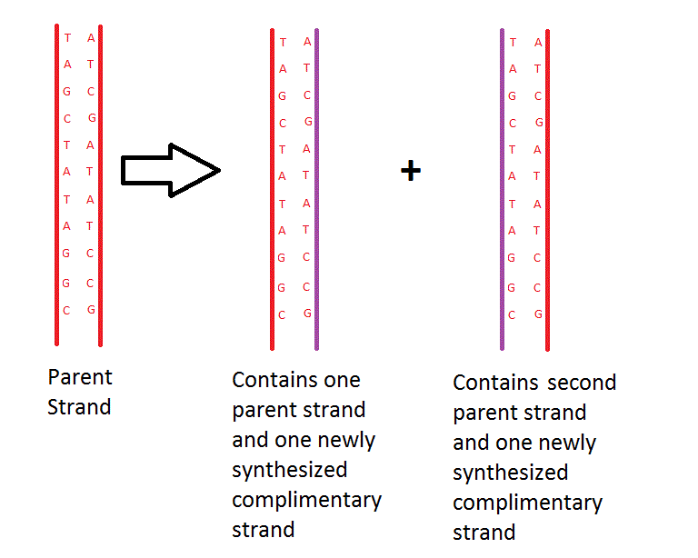
| 11:34, 17 November 2011 | Semi-conservative replication.gif (file) |  |
20 KB | 110365880 (talk | contribs) | 1 | |
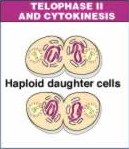
| 20:44, 14 November 2011 | Telophase II meiosis.png (file) |  |
31 KB | 110029052 (talk | contribs) | A diagram showing telophase in the second stage of cell division in meiosis (meiosis II. | 1 |
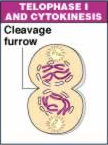
| 20:43, 14 November 2011 | Telophase I meiosis.png (file) |  |
26 KB | 110029052 (talk | contribs) | A diagram showing telophase in the first cell division stage of meiosis (meiosis I. | 1 |
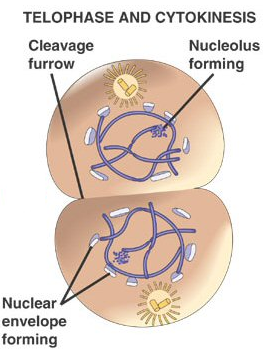
| 20:42, 14 November 2011 | Telophase mitosis.png (file) |  |
122 KB | 110029052 (talk | contribs) | A diagram showing the last stage of mitosis known as telophase. | 1 |
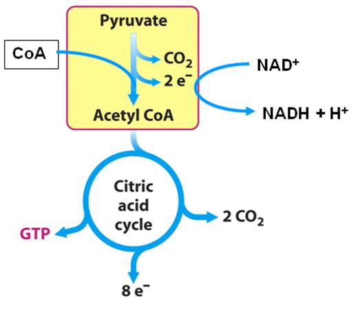
| 18:33, 14 November 2011 | Pyruvate+CAC.jpg (file) |  |
21 KB | 104097300 (talk | contribs) | Step 0 and Cycle as Steps 1-8 | 1 |
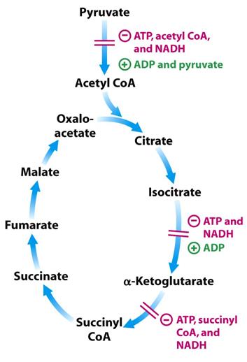
| 18:32, 14 November 2011 | Control of CAC.jpg (file) |  |
21 KB | 104097300 (talk | contribs) | Control of the Krebs cycle by catalysis of irreversible steps | 1 |
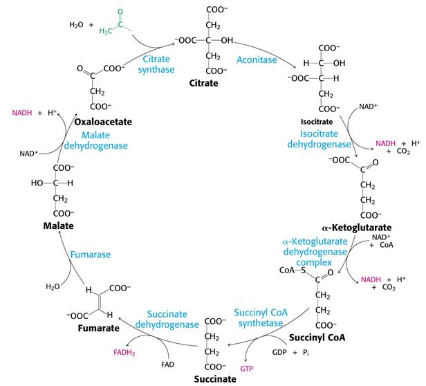
| 18:30, 14 November 2011 | CAC detail.jpg (file) |  |
34 KB | 104097300 (talk | contribs) | Krebs cycle in detail | 1 |
| 18:30, 14 November 2011 | Anabolic properties of CAC.jpg (file) |  |
22 KB | 104097300 (talk | contribs) | Anabolic properties of the Krebs Cycle | 1 |
| 16:25, 14 November 2011 | Aspartic acid.gif (file) |  |
6 KB | 100559770 (talk | contribs) | 3D Structure of aspartic acid | 1 |
| 15:39, 14 November 2011 | Aromatic ring for wiki.gif (file) |  |
2 KB | 100240801 (talk | contribs) | Reverted to version as of 15:34, 14 November 2011 | 3 |
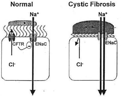
| 15:23, 14 November 2011 | Disease and treatment 2.jpg (file) |  |
19 KB | 104097300 (talk | contribs) | Effect of cystic fibrosis on ENaC | 1 |
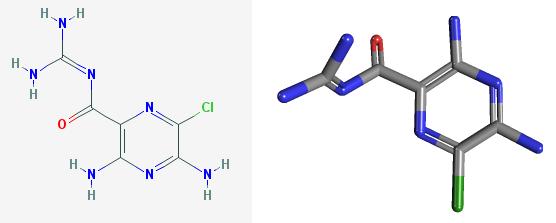
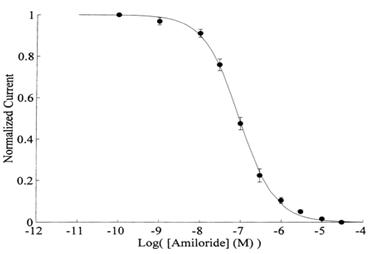
| 15:22, 14 November 2011 | Disease and treatment 1-amiloride from pubchem CID 2016231.jpg (file) |  |
11 KB | 104097300 (talk | contribs) | 2D and 3D structure of amiloride | 1 |
| 15:21, 14 November 2011 | Regulation 5.jpg (file) |  |
10 KB | 104097300 (talk | contribs) | Removal and insertion of ENaC | 1 |
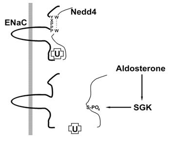
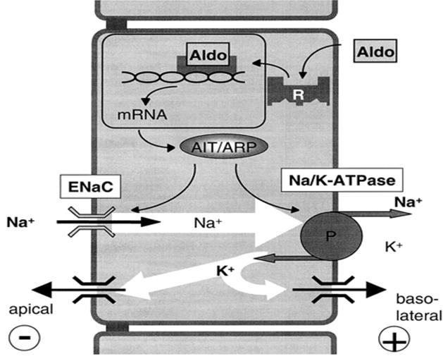
| 15:20, 14 November 2011 | Regulation 4.jpg (file) |  |
42 KB | 104097300 (talk | contribs) | Effect of aldosterone on ENaC | 1 |
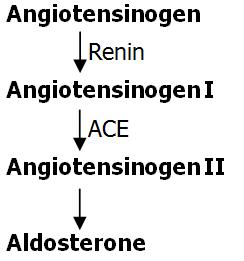
| 15:20, 14 November 2011 | Regulation 3 (RAAS).jpg (file) | .jpg) |
12 KB | 104097300 (talk | contribs) | Renin-Angiotensin-Aldosterone System | 1 |
| 15:19, 14 November 2011 | Regulation 2.jpg (file) |  |
69 KB | 104097300 (talk | contribs) | Activation of ENaC via protease cleaving | 1 |
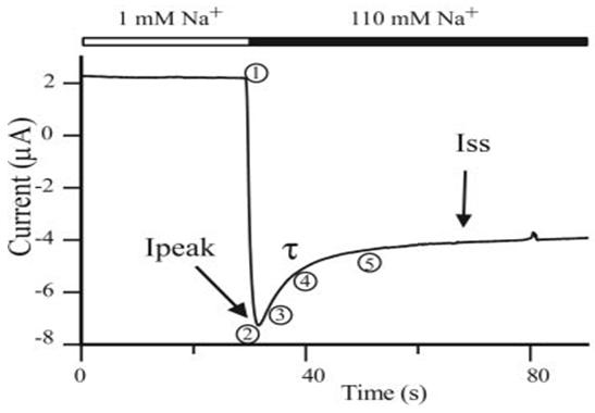
| 15:18, 14 November 2011 | Regulation 1.jpg (file) |  |
16 KB | 104097300 (talk | contribs) | How ENaC responds to increased extracellular Na+ increase | 1 |
| 15:17, 14 November 2011 | Structure 5.jpg (file) |  |
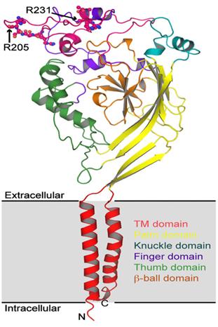
12 KB | 104097300 (talk | contribs) | ENaC C & N-termini, cysteine rich domains of extracellular loop and 2TMs | 1 |
| 15:16, 14 November 2011 | Structure 4.jpg (file) |  |
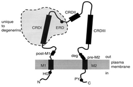
29 KB | 104097300 (talk | contribs) | ENaC domains | 1 |
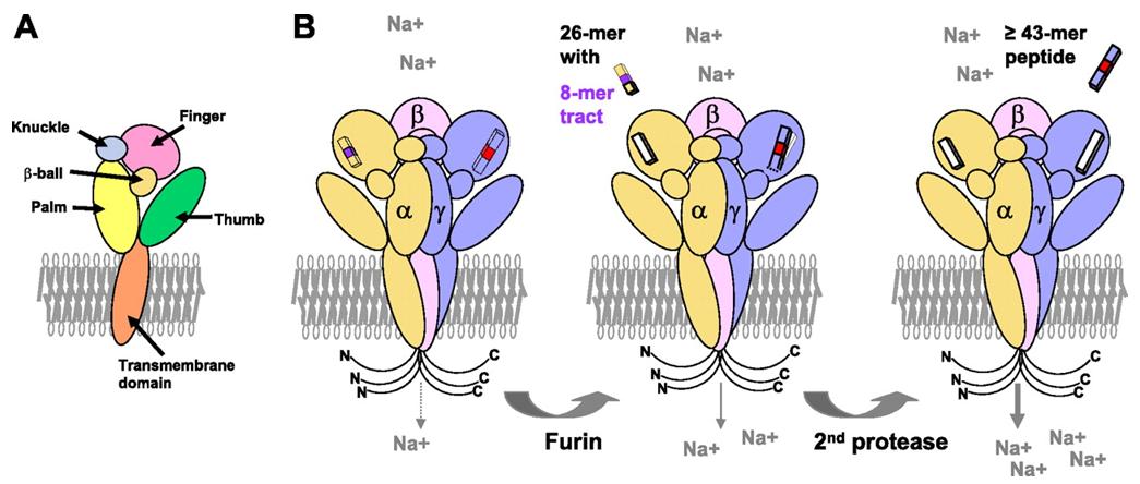
| 15:15, 14 November 2011 | Structure 3.jpg (file) |  |
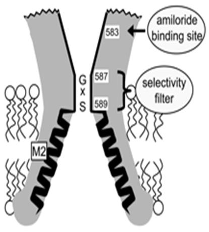
22 KB | 104097300 (talk | contribs) | cartoon of ENaC structure and selectivity filter | 1 |
| 15:14, 14 November 2011 | Structure 2.jpg (file) |  |
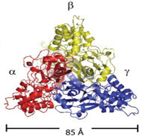
29 KB | 104097300 (talk | contribs) | ENaC trimeric structure | 1 |
| 15:14, 14 November 2011 | Structure 1.jpg (file) |  |
7 KB | 104097300 (talk | contribs) | alpha, beta and gamma subunits must be co-expressed | 1 |
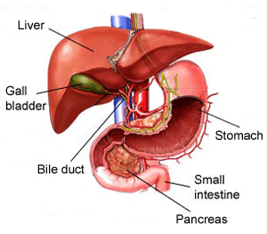
| 14:24, 11 November 2011 | Liver diagram.gif (file) |  |
31 KB | 100560053 (talk | contribs) | Liver drawing | 1 |
| 10:29, 11 November 2011 | Mmdbimage.jpg (file) |  |
8 KB | 100234637 (talk | contribs) | 1 | |
| 11:03, 10 November 2011 | Protein structure.png (file) |  |
165 KB | 100431328 (talk | contribs) | Protein showing hydrophobic and hydrophilic regions | 1 |
| 11:01, 10 November 2011 | Table.jpg (file) |  |
30 KB | 100431328 (talk | contribs) | Table of polar amino acids | 1 |