Uncategorized files
Jump to navigation
Jump to search
Showing below up to 50 results in range #301 to #350.
-
 Structure 2.jpg 495 × 473; 29 KB
-
 Structure 3.jpg 414 × 459; 22 KB
-
 Structure 4.jpg 311 × 466; 29 KB
-
 Structure 5.jpg 434 × 284; 12 KB
-
 Structure of Glucokinase.jpg 500 × 500; 17 KB
-
 Structure of oseltamivir.png 200 × 200; 5 KB
-
 Sugar Phosphate Backbone.png 252 × 263; 13 KB
-
 Sulphur Lumps 99 98 .jpg 1,024 × 768; 88 KB
-
 Synapse.jpg 569 × 525; 102 KB
-
 Table.jpg 465 × 199; 30 KB
-
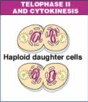 Telophase II meiosis.png 129 × 149; 31 KB
-
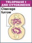 Telophase I meiosis.png 108 × 145; 26 KB
-
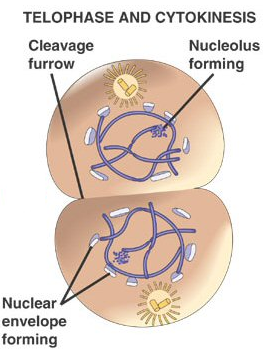 Telophase mitosis.png 279 × 352; 122 KB
-
 Tertiary.jpg 1,117 × 637; 287 KB
-
 Test.png 141 × 185; 38 KB
-
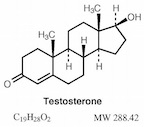 Testosterone Molecule.jpg 144 × 127; 12 KB
-
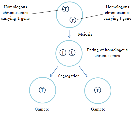 The Principle of Segregation (diagram).png 540 × 460; 17 KB
-
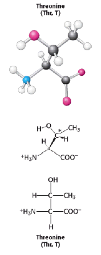 Threonine.png 201 × 505; 40 KB
-
 Tight junction image.png 544 × 451; 221 KB
-
 Tp11 02.jpg 450 × 338; 24 KB
-
 Transcription for WIKI.png 413 × 500; 43 KB
-
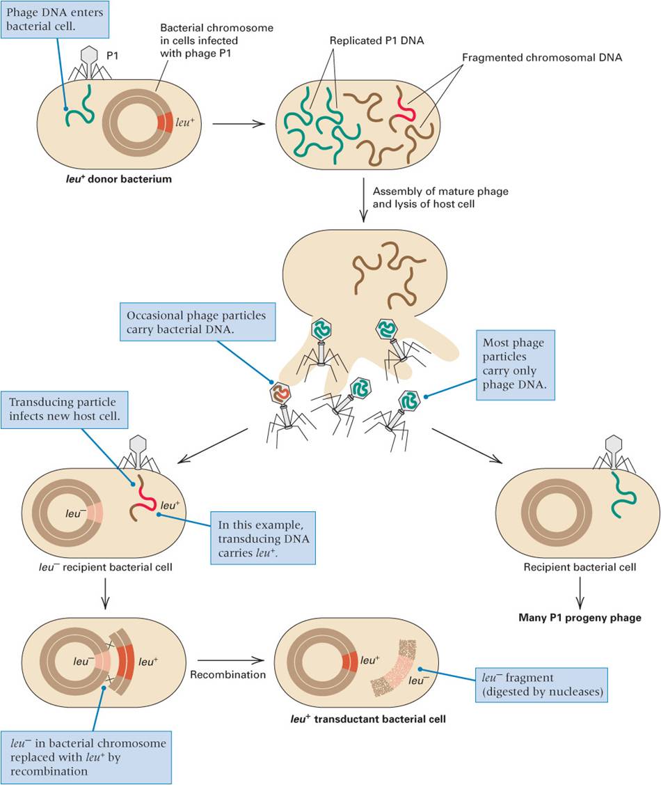 Transduction.jpg 950 × 1,127; 96 KB
-
 Triple X karyotype.gif 440 × 410; 25 KB
-
 Tripple =.png 8 × 7; 129 bytes
-
 Trisomy13.jpg 275 × 360; 24 KB
-
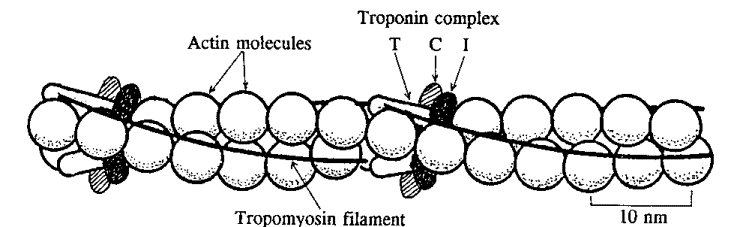 Tropomyosin.gif 752 × 227; 18 KB
-
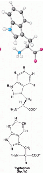 Tryptophan.png 160 × 594; 82 KB
-
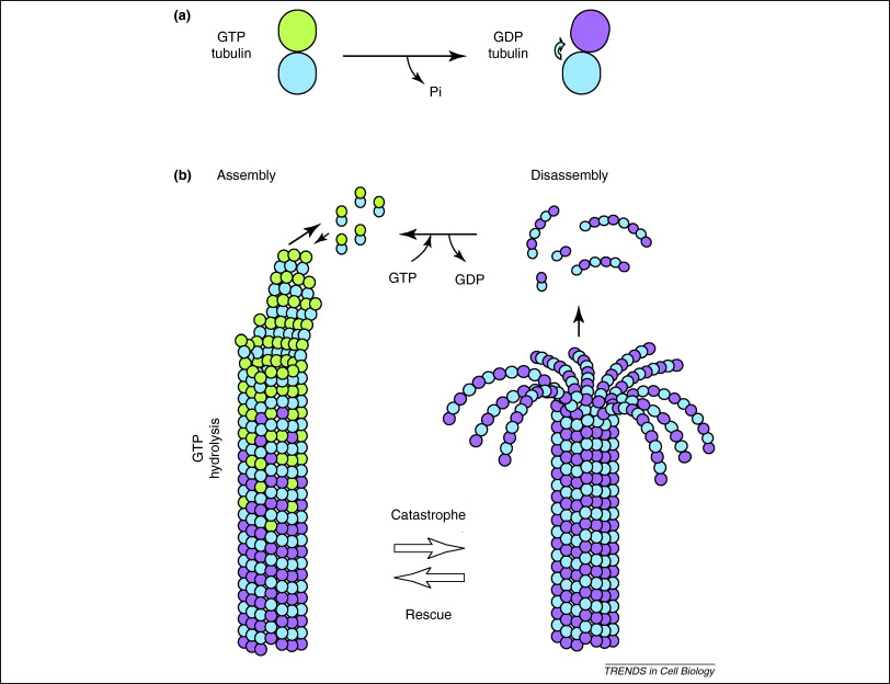 Tubulin.jpg 812 × 624; 84 KB
-
 Types of summation.jpg 427 × 400; 43 KB
-
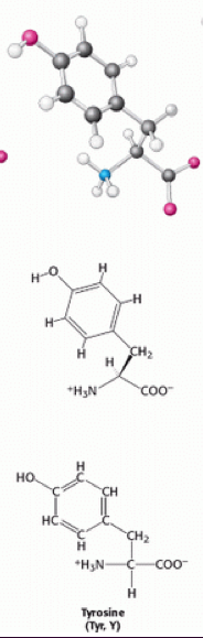 Tyrosine.png 184 × 579; 82 KB
-
 UNADJUSTEDNONRAW thumb 38a.jpg 550 × 353; 33 KB
-
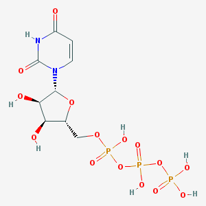 UTP.png 300 × 300; 4 KB
-
 UTP3D.png 300 × 300; 23 KB
-
 Untitled.png 614 × 260; 71 KB
-
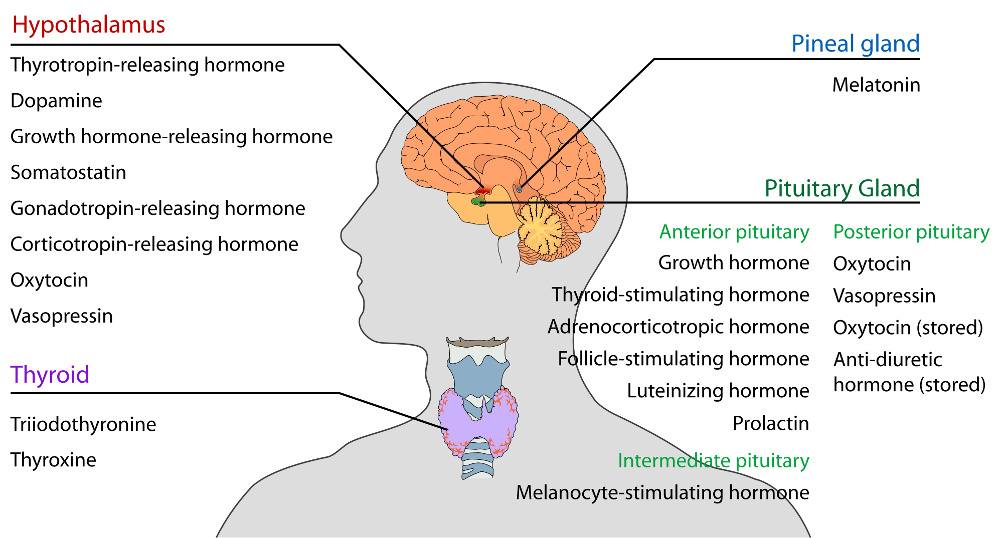 Upper-Endocrine-Glands.png 2,000 × 1,074; 391 KB
-
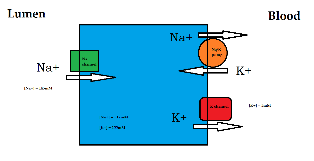 Ussing model.png 1,260 × 646; 28 KB
-
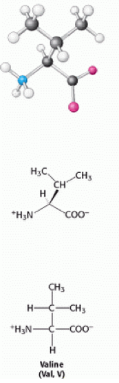 Valine.png 173 × 556; 60 KB
-
 Vein med.jpg 580 × 360; 40 KB
-
 Visible Spectrum.png 466 × 148; 16 KB
-
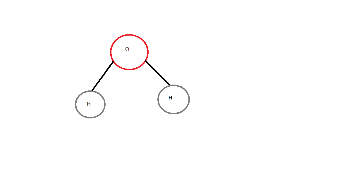 WATER MOLECULE.png 1,152 × 648; 8 KB
-
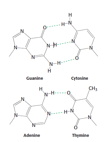 Watson-Crick Base Pairs.PNG 361 × 470; 18 KB
-
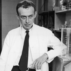 Watson.jpg 300 × 300; 14 KB
-
 Watson n crick.jpg 196 × 146; 7 KB
-
 Watson postcard.jpg 280 × 396; 62 KB
-
 Wiki.jpg 457 × 100; 13 KB
-
 Wiki pic.jpg 720 × 540; 35 KB
-
 Working principle of telomerase.png 400 × 535; 154 KB
-
 Wow.png 350 × 186; 4 KB
-
 X-ray.jpg 145 × 144; 4 KB
-
 X chromosome.jpg 390 × 183; 12 KB


.png)


