DNA

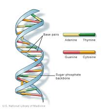
DNA (deoxyribonucleic acid) is the genetic information found in the nuclei of most organisms. It is arranged into structures called chromosomes. The structure of DNA was first identified as having a 'double-helix' structure by Watson and Crick in 1953. DNA is composed of 4 bases: the purines, adenine (A) and guanine (G) and pyrimidines ,thymine (T) and cytosine (C)[1]. These form complementary base pairs of A-T and G-C. DNA also contains a phosphate group connected to a deoxyribose sugar. The phosphate group is attached to the sugar through a phosphodiester bond. Humans have 99.5% similarities with other humans in their DNA.
Structure of DNA
DNA (deoxyribonucleic acid) is a chain of monomers (repeating units) called "nucleotides". A nucleotide consists of: a 2` deoxyribose sugar (A five-carbon pentose similar to that of ribose sugar found in RNA. Its chemical formula is C5H10O4), a phosphate group (which forms a phosphodiester bond: connecting 2 deoxyribose sugars together) and a nitrogenous base (one from A (Adenine), C ( Cytosine), G ( Guanine) or T (Thymine), which forms a side chain branching from the 1' carbon of the 2` deoxyribose sugar).
The deoxyribose sugar/phosphate group region is regarded as the 'backbone' of DNA strands due to its structural purpose and the sequence of bases carries the gentic information. In order to produce a double-stranded DNA structure, interactions occur between complementary bases. The complementary base pairs in DNA interact with one another via hydrogen bonds: A-T interactions consist of 2 intermolecular hydrogen bonds, whereas G-C interactions consist of 3 intermolecular hydrogen bonds. In between these bases are hydrophobic interactions known as van der Waal forces[2]. These interactions form bridges between two DNA chains, thus creating a double-stranded 'ladder' shaped structure. Each strand acts as a template for the other one in DNA replication. DNA is copied into mRNA (messenger RNA) which carries the information from the original DNA template strand to be involved in protein synthesis. The process of DNA being copied into mRNA is termed transcription. The transcribed mRNA is then translated to a polypeptide in a process called translation by tRNA.
In the DNA double helix the strands of the backbone are closer together on one side of the helix than they are on the other. This leads to the formation of major and minor grooves[3]. The major groove is much wider than the minor groove and this means that specific DNA-protein interactions can take place on the major groove due to the backbone not being in the way. The specific nucleotides that face into the major groove are the N7 and C6 groups of purines and the C4 and C5 groups of pyrimidines, which accept hydrogen ions from the amino acids in the protein to form hydrogen bonds[4].
Due to the double helical structure of DNA, the nitrogenous bases are found on the inside of the structure, forming a hydrophobic interior. The negative charge from the phosphate groups gives the sugar-phosphate backbone of DNA a negative charge, which repels nucleophiles, including water. This makes DNA less vulnerable to nucleophilic attack, thus DNA is considered to be a very stable molecule. DNA is much more stable then RNA since RNA is only single-stranded - the nitrogenous bases are left exposed to attack by nucleophiles on one side.
In 1953, despite many other theories, James Watson and Francis Crick discovered the true structure of a double stranded DNA molecule to be a 'Double Helix'. This was solved as a result of 'stick-and-ball' models they created, along with utilising the work of fellow scientists Rosalind Franklin and Maurice Wilkins on X-ray crystallography[5]. The X-ray diffraction photographs obtained from DNA fibres, displayed a unique X-shape, which illustrates a helical stucture, although they indicated a repeating structure of 3.4 Å apart per turn of the helix, each base is rotated 36 degrees from the next one. The diameter of the helix is 23.7 Å. They found that the sugar-phosphate backbone was on the outside and the bases are positioned on the inside of the helix[6][7].
The above information described the B-form of DNA. DNA is also found in A- and Z-forms[8]. When the DNA becomes dehydrated, the A-form can be observed[9]. It is also right-handed, but there are 11 bases per turn and the helix is broader. The diameter is 25.5 Å. Another difference is that the tilt of the base pairs increases by 18o, to 19o from perpendicular to the helix axis.
The Z-form differs far more as it is a left-handed double helix. This form is rarely seen without the help of high salt concentrations[10]. The bonds are zigzagged as the bonds are alternating anti and syn (whereas A- and B-forms are anti only). The Z-form is narrower, having a diameter of only 18.4 Å, but there is a 3.8 Å rise per base pair. It is thought that transitions between the B and Z forms of DNA may be involved in the regulation of gene regulation.
B-DNA is most commonly seen in all life forms[11], however, A-helical and Z-helical structures co-exsist in cells; i.e. it is very common to observe a molecule of B-DNA and Z-DNA , in a predominantly B-DNA confirmation.
The DNA of the Indian muntjac which is an Asiatic deer has the longest length (approximately 3 billion nucleotides) among all the known DNA molecules of other organisms[3].
DNA is negatively charged due to the negativley charged phosphate ions in the sugar-phosphate backbone. Hence it can be used for gel electrophoresis to identify different lengths of DNA. The negative charge of the backbone, along with the OH-groups on the deoxyribose sugar, means that the backbone is Hydrophillic as water can form hydrogen bonds with it. The centre of the DNA molecule is hydrophobic due to the lack of charge in DNA bases. The hydrophillic outer and hydrophobic inner of the DNA molecule means that it is soluble in water[12].
Replication
The double stranded nature of DNA is important to the "semi-conservative replication" method of DNA replication. In this process, the enzyme DNA helicase unwinds the double helix by breaking the hydrogen bonds between the complementary bases on each strand revealing the 2 seperate strands. On these strands are the revealed bases, which attract complementary bases on free nucleotides. The free nucleotides are joined together by an enzyme DNA polymerase. Deoxyribonucleotide triphosphate (dNTPs) are added onto the 3' hydroxyl group on the growing strand through the 5' triphosphate group on the incoming dNTP in a esterification reaction[13]. The joining of nucleotides forms a new strand of DNA which is identical to the other double strand of DNA, as it uses one of the original strands as a template for replication. Each daughter double strand of DNA is made up of a parent strand and a newly sythesised strand.
Even though both strands in the parental DNA molecule are copied to form identical products, the two strands are copied in a slightly different manner from each other. This is due to the fact that DNA is always synthesised in a 5' to 3' direction. The 3' to 5' strand, known as the leading strand, is copied continuously by DNA polymerase. The other strand is called the lagging strand, as it is replicated more slowly. To replicate the lagging strand, RNA primers are placed on several points along the lagging strand by an enzyme called primase. The gaps on the lagging strand between the RNA primers are replicated by DNA polymerase, and the short fragments of replicated DNA are known as Okazaki fragments. However, in order to complete the replication of the lagging strand, RNA primers must be replaced by DNA sequences. Another DNA polymerase removes the RNA primers and synthesises DNA fragments to replace them. The Okazaki fragments and the RNA primer replacements are still not joined, so DNA ligase comes in an ligates all the fragments of DNA together[14].
The theory of semi-conservative replication was proven to be correct by the Messelson-Stahl experiment. In this experiment, E.coli were grown in a medium containing 15-N for a number of generations. The bacteria were then transferred to a medium containing 14-N. After one replication cycle DNA was extracted from the bacteria and centrifuged. The centrifugation separated the DNA by density, producing one band wita h density between that of 15-N DNA and 14-N DNA. This showed that one strand came from the parent (15-N) and one strand was newly synthesised from free nucleotides (14-N)[15].
References
- ↑ HARTL AND JONES,2009:41, GENETICS : ANALYSIS OF GENES AND GENOMES SEVENTH EDITION.
- ↑ Lawrie Ryan and Roger Norris, Cambridge International AS and A Level, Second Edition, Cambridge United Kingdom, Latimer Trend 2014
- ↑ 3.0 3.1 Boston University (N.d.) 'Major and Minor Grooves', Available at: https://tandem.bu.edu/knex/knex.pdf Cite error: Invalid
<ref>tag; name "null" defined multiple times with different content - ↑ Delmar Larsen (N.d.), 'B-Form, A-Form, Z-Form of DNA', Available at: http://biowiki.ucdavis.edu/Genetics/Unit_I%3A_Genes,_Nucleic_Acids,_Genomes_and_Chromosomes/Chapter_2._Structures_of_nucleic_acids/B-Form,_A-Form,_Z-Form_of_DNA, Accessed: 25th November 2014
- ↑ http://nobelprize.org/educational/medicine/dna_double_helix/readmore.html
- ↑ http://www.chm.bris.ac.uk/motm/dna/dna.htm
- ↑ J.Berg, J.Tymoczko, L.Stryer;, 113-115, 2012 Freeman; Biochemistry
- ↑ Berg, J.M., Tymoczko, J.L. and Stryer, L. (2011) Biochemistry. Seventh Edition. Basingstoke: Basingstoke : Palgrave Macmillan. p20
- ↑ Ferrier, D.R. (2014) Biochemistry. Baltimore, Md. ; London: Baltimore, Md. ; London : Lippincott Williams and Wilkins. p398
- ↑ Bae, S., Kim, D., Kim, K.K., Kim, Y.-g., Hohng, S. and Kim, Y.-G. (2011) 'Intrinsic Z- DNA is stabilized by the conformational selection mechanism of Z- DNA-binding proteins', Journal of the American Chemical Society, 133(4), p. 668.
- ↑ Malacinski, G. Essentials of molecular biology.4th Ed. New Delhi: Narosa. 2003.
- ↑ Ruvolo M, Hartl, DL (2012) Genetics : analysis of genes and genomes, 8th ed, Burlington, MA : Jones and Bartlett Learning
- ↑ Berg JM, Tymoczko JL, Gatto GJ jr, Stryer L. Biochemistry. 8th ed. New York: W.H. Freeman and Company. 2015. P107-111
- ↑ Hartl, D. L. and Ruvolo, M., 2012. Genetics: Analysis of Genes and Genomes. 8th ed. Burlington: Jones and; Bartlett Learning. Pages 205-210
- ↑ Berg J.M, Tymoczko J.L, Stryer L, Biochemistry, 7th ed. 2012:123, New York: W.H Freeman and Company