The Cardiovascular System: Difference between revisions
No edit summary |
No edit summary |
||
| Line 1: | Line 1: | ||
The Cardiovascular System (also called the circulatory system) circulates blood around the body in order to supply all tissue with the required nutrients and [[Image:Cardiovascularsystem.jpg|frame|right]][[Oxygen|oxygen]] and remove waste products such as [[Carbon dioxide|carbon dioxide]]<ref name="parker"> Parker S. (2007) The Human Body Book, 1st edition, London: Dorling Kindersley Limited.</ref>. Consequently it is crucial for [[Homeostasis|homeostasis]]<ref name="website">http://stayhealthier.wordpress.com/2010/03/30/cardiovascular-system-understand-it-to-stay-healthy/</ref>. | The Cardiovascular System (also called the circulatory system) circulates blood around the body in order to supply all tissue with the required nutrients and [[Image:Cardiovascularsystem.jpg|frame|right|Cardiovascularsystem.jpg]][[Oxygen|oxygen]] and remove waste products such as [[Carbon dioxide|carbon dioxide]]<ref name="parker"> Parker S. (2007) The Human Body Book, 1st edition, London: Dorling Kindersley Limited.</ref>. Consequently it is crucial for [[Homeostasis|homeostasis]]<ref name="website">http://stayhealthier.wordpress.com/2010/03/30/cardiovascular-system-understand-it-to-stay-healthy/</ref>. | ||
In order to carry out its functions, the cardiovascular system pumps blood through the blood vessels. This is done by the pumping of the heart. Furthermore, the circulatory system is associated with other systems in the body, for example, the lymphatic system<ref name="parker" />. | In order to carry out its functions, the cardiovascular system pumps blood through the blood vessels. This is done by the pumping of the heart. Furthermore, the circulatory system is associated with other systems in the body, for example, the lymphatic system<ref name="parker" />. | ||
| Line 9: | Line 9: | ||
Within the human body, blood is carried in blood vessels which can differ in size and makeup<ref name="parker" />. | Within the human body, blood is carried in blood vessels which can differ in size and makeup<ref name="parker" />. | ||
===== [ | ===== [[Arteries|Arteries]] ===== | ||
Initiate from the heart and carry blood to organs and tissues where [[Oyxgen|oyxgen]] and nutrients are needed. Therefore, they carry oxygenated blood away from the heart to the tissues. The diameter of the larger arteries can be altered by smooth muscle contraction and relaxation in order to maintain blood pressure, keeping the blood flowing. The smooth muscle in the walls of the arteries are controlled by the [[Autonomic Nervous System|autonomic nervous system ]](ANS), which is part of the [[Peripheral Nervous System|peripheral nervous system]]. The ANS acts unconsciously on the smooth muscle, allowing rapid contraction/dilation of the vessels in response to many different factors such as blood pressure and heart rate. Thick walls, muscular (see [[Cardiac muscle|cardiac muscle]]), and elastic layers ensure that the strain due to the blood flow and the contraction of the heart does not damage the walls, which on average are only 1 mm thick<ref name="parker" />. | Initiate from the heart and carry blood to organs and tissues where [[Oyxgen|oyxgen]] and nutrients are needed. Therefore, they carry oxygenated blood away from the heart to the tissues. The diameter of the larger arteries can be altered by smooth muscle contraction and relaxation in order to maintain blood pressure, keeping the blood flowing. The smooth muscle in the walls of the arteries are controlled by the [[Autonomic Nervous System|autonomic nervous system ]](ANS), which is part of the [[Peripheral Nervous System|peripheral nervous system]]. The ANS acts unconsciously on the smooth muscle, allowing rapid contraction/dilation of the vessels in response to many different factors such as blood pressure and heart rate. Thick walls, muscular (see [[Cardiac muscle|cardiac muscle]]), and elastic layers ensure that the strain due to the blood flow and the contraction of the heart does not damage the walls, which on average are only 1 mm thick<ref name="parker" />. | ||
===== [ | ===== [[Veins|Veins]] ===== | ||
In contrast to the arteries, veins have thinner walls and are therefore more flexible. These properties arise from the low blood pressure within veins, causing the blood flow to be much slower. A further characteristic of veins are the valves they contain, which are made from endothelium tissue and restrict the blood flow in one direction<ref name="parker" />. Valves contain of two cusps that only open into the direction of blood flow due to pumping of the [[Heart|heart]]. | In contrast to the arteries, veins have thinner walls and are therefore more flexible. These properties arise from the low blood pressure within veins, causing the blood flow to be much slower. A further characteristic of veins are the valves they contain, which are made from endothelium tissue and restrict the blood flow in one direction<ref name="parker" />. Valves contain of two cusps that only open into the direction of blood flow due to pumping of the [[Heart|heart]]. | ||
| Line 19: | Line 19: | ||
This is especially important regarding their function, as this is to return deoxygenated blood to the heart. The main two veins doing this are called superior and inferior venae cavae<ref name="parker" />. | This is especially important regarding their function, as this is to return deoxygenated blood to the heart. The main two veins doing this are called superior and inferior venae cavae<ref name="parker" />. | ||
===== [ | ===== [[Capillaries|Capillaries]] ===== | ||
As the distance of the blood vessels to the heart increases, the amount of branching increases and the blood vessels become smaller and finer in order for the blood to reach every part of the body. Starting with large arteries, these blood vessels then further divide into little vessels called capillaries (0.01 mm in diameter). Not only the size decreases but also the thickness of the walls decreases to a layer of only one cell so that a maximum exchange of gases with cells and [[Tissue|tissues]] can be achieved<ref name="parker" />. | As the distance of the blood vessels to the heart increases, the amount of branching increases and the blood vessels become smaller and finer in order for the blood to reach every part of the body. Starting with large arteries, these blood vessels then further divide into little vessels called capillaries (0.01 mm in diameter). Not only the size decreases but also the thickness of the walls decreases to a layer of only one cell so that a maximum exchange of gases with cells and [[Tissue|tissues]] can be achieved<ref name="parker" />. | ||
| Line 43: | Line 43: | ||
CHD includes common conditions like [[Atherosclerosis|atherosclerosis]], [[Angina|angina]] or [[Heart attack|Heart attack]]<ref name="parker" />. | CHD includes common conditions like [[Atherosclerosis|atherosclerosis]], [[Angina|angina]] or [[Heart attack|Heart attack]]<ref name="parker" />. | ||
===== [[Atherosclerosis|Atherosclerosis]] ===== | ===== [[Atherosclerosis|Atherosclerosis]] ===== | ||
This disease is due to fatty deposits that lign and eventually clog the arteries, causing them to narrow and become inflexible. Sources of the fatty deposits are excess fat and [[Cholesterol|cholesterol]] in the blood, which settles on the artery walls as the blood flows through. These deposits are then known as atheroma. These structures in turn form plaques as they start to accumulate. As these plaques build up, the lumen of the diseased artery narrows and consequently blood flow is restricted. In fatal cases, blood flow is not possible beyond the plaque, so blood supply becomes insufficient and body parts like the [[Brain|brain]] may suffer oxygen starvation can result in a [[Stroke|stroke]] (also see [[Angina|Angina]])<ref name="parker" />. | This disease is due to fatty deposits that lign and eventually clog the arteries, causing them to narrow and become inflexible. Sources of the fatty deposits are excess fat and [[Cholesterol|cholesterol]] in the blood, which settles on the artery walls as the blood flows through. These deposits are then known as atheroma. These structures in turn form plaques as they start to accumulate. As these plaques build up, the lumen of the diseased artery narrows and consequently blood flow is restricted. In fatal cases, blood flow is not possible beyond the plaque, so blood supply becomes insufficient and body parts like the [[Brain|brain]] may suffer oxygen starvation can result in a [[Stroke|stroke]] (also see [[Angina|Angina]])<ref name="parker" />. | ||
Revision as of 17:49, 18 October 2018
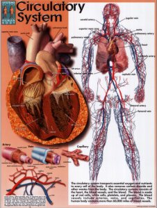
The Cardiovascular System (also called the circulatory system) circulates blood around the body in order to supply all tissue with the required nutrients and

oxygen and remove waste products such as carbon dioxide[1]. Consequently it is crucial for homeostasis[2].
In order to carry out its functions, the cardiovascular system pumps blood through the blood vessels. This is done by the pumping of the heart. Furthermore, the circulatory system is associated with other systems in the body, for example, the lymphatic system[1].
Blood and Blood Vessels
Blood consists of white blood cells, red blood cells, platelets and liquid plasma.Plasma accounts for about 50% of the total blood content and consists mainly of which has glucose, proteins such as enzymesand hormones and waste products dissolved in it. Oxygen and carbon dioxide are also present in the blood and are carried by the red blood cells[1].
Within the human body, blood is carried in blood vessels which can differ in size and makeup[1].
Initiate from the heart and carry blood to organs and tissues where oyxgen and nutrients are needed. Therefore, they carry oxygenated blood away from the heart to the tissues. The diameter of the larger arteries can be altered by smooth muscle contraction and relaxation in order to maintain blood pressure, keeping the blood flowing. The smooth muscle in the walls of the arteries are controlled by the autonomic nervous system (ANS), which is part of the peripheral nervous system. The ANS acts unconsciously on the smooth muscle, allowing rapid contraction/dilation of the vessels in response to many different factors such as blood pressure and heart rate. Thick walls, muscular (see cardiac muscle), and elastic layers ensure that the strain due to the blood flow and the contraction of the heart does not damage the walls, which on average are only 1 mm thick[1].
In contrast to the arteries, veins have thinner walls and are therefore more flexible. These properties arise from the low blood pressure within veins, causing the blood flow to be much slower. A further characteristic of veins are the valves they contain, which are made from endothelium tissue and restrict the blood flow in one direction[1]. Valves contain of two cusps that only open into the direction of blood flow due to pumping of the heart.
This is especially important regarding their function, as this is to return deoxygenated blood to the heart. The main two veins doing this are called superior and inferior venae cavae[1].
As the distance of the blood vessels to the heart increases, the amount of branching increases and the blood vessels become smaller and finer in order for the blood to reach every part of the body. Starting with large arteries, these blood vessels then further divide into little vessels called capillaries (0.01 mm in diameter). Not only the size decreases but also the thickness of the walls decreases to a layer of only one cell so that a maximum exchange of gases with cells and tissues can be achieved[1].
The capillaries carrying waste products away from the cells and tissues eventually enlarge into veins which carry the blood back to the heart[1].
Heart Structure
The wall of the heart consists of myogenic muscle which is called myocardium. Just like all other tissues in the body, this muscle requires nutrients and oxygen which are supplied by blood vessels called coronary arteries. These originate from the right and the left artery which in turn divide from the aorta. Naturally, waste products must be removed as well which is done by the coronary sinus[1].
Inside the heart, hollow chambers can be found which are filled with blood and emptied again during each contraction cycle. The top chambers are called right and left atrium and receive blood from the body. They are located above the right and left ventricle respectively which pump blood into the arteries to the body. The upper and lower chambers are separated by valves and the right and left side are separated by the septum[1]. Valves allow blood to flow into the ventricles from the system during diastole and again closes during ventricular systole in order to avoid the blood flowing back when the heart contracts[3].
The muscle wall of the left-hand side of the heart is much thicker and stronger due to the fact that it has to pump blood into the whole system that supplies the body with blood and therefore has to supply more powerful contraction.
(see also Heart)
In order to control the contraction of all the different regions of the heart, there is also a network of ordered nerves and nodes. The sinoatrial node which is situated above the right atrium, constantly generates signals that travel through the right atrium by the atrioventricular node, causing a contraction. The signal then travels down the septum, along the sides of both ventricles via the bundle of hiss, into the bottom the heart. Here, this wave of excitation races through purkinje fibres up the walls of the ventricles, which causes a contraction. This arrangement of nerves means ventricles contract after the atriums.
This type of disease is caused by insufficient nutrient and oxygen supply to the heart muscle as a consequence of damaged or malfunctioning coronary arteries[1].
CHD includes common conditions like atherosclerosis, angina or Heart attack[1].
This disease is due to fatty deposits that lign and eventually clog the arteries, causing them to narrow and become inflexible. Sources of the fatty deposits are excess fat and cholesterol in the blood, which settles on the artery walls as the blood flows through. These deposits are then known as atheroma. These structures in turn form plaques as they start to accumulate. As these plaques build up, the lumen of the diseased artery narrows and consequently blood flow is restricted. In fatal cases, blood flow is not possible beyond the plaque, so blood supply becomes insufficient and body parts like the brain may suffer oxygen starvation can result in a stroke (also see Angina)[1].
Major factors that influence fatty deposits are smoking, saturated fat-rich diet, lack of exercise and overweight[1].
Angina
Angina is a condition that can also be described as chest pain and results from insufficient blood supply to the heart muscle. This insufficient blood supply means the heart muscle lacks oxygen, which in turn stresses the heart when it works at an increased rate. This might occur during exercise, stress, cold weather or large food intake but can be relieved by relaxation and subsequent decrease in the rate of activity of the heart muscle. As explained in the above section, the insufficient blood supply may result from atherosclerosis where the arteries are narrowed[1].
Due to oxygen starvation, the heart muscle may be damaged over time (see Heart Attack)[1].
Heart Attack
This type of condition is also called Myocardial Infarction and results from oxygen starvation in an area of the heart muscle where blood flow was restricted or lacking. The restricted blood flow results from blockage or narrowing of the arteries in the heart muscle (as described in Atherosclerosis) and leads to the formation of a blood clot or thrombus. The lack of oxygen and the accumulation of waste products then causes tissue death within this specific area of the cardiac muscle[1].
Symptoms of a heart attack are sweating, shortness of breath, nausea and loss of consciousness[1].
References
- ↑ 1.00 1.01 1.02 1.03 1.04 1.05 1.06 1.07 1.08 1.09 1.10 1.11 1.12 1.13 1.14 1.15 1.16 1.17 1.18 Parker S. (2007) The Human Body Book, 1st edition, London: Dorling Kindersley Limited.
- ↑ http://stayhealthier.wordpress.com/2010/03/30/cardiovascular-system-understand-it-to-stay-healthy/
- ↑ Brown H. (1997) Physiology and Pharmacology of the Heart, 1st edition, Oxford: Blackwell Science Limited.