Muscle: Difference between revisions
No edit summary |
No edit summary |
||
| Line 1: | Line 1: | ||
== Skeletal Muscle<br> == | == Skeletal Muscle<br> == | ||
See [[Skeletal Muscle|Skeletal Muscle]] | |||
[[ | |||
Skeletal | |||
== Cardiac Muscle == | == Cardiac Muscle == | ||
| Line 39: | Line 13: | ||
== Smooth Muscle == | == Smooth Muscle == | ||
Smooth muscle tissue is classed as non-striated due it's appearance and cells are located mainly in the walls of hollow organs such as the urinary, reproductive, intestinal and respiratory tracts of both females and males.<ref>http://books.google.se/books?id=iOEQWGfiurYC&amp;amp;amp;amp;amp;amp;amp;amp;amp;amp;amp;amp;amp;pg=PA175&amp;amp;amp;amp;amp;amp;amp;amp;amp;amp;amp;amp;amp;lpg=PA174#v=onepage&amp;amp;amp;amp;amp;amp;amp;amp;amp;amp;amp;amp;amp;q&amp;amp;amp;amp;amp;amp;amp;amp;amp;amp;amp;amp;amp;f=false</ref> contraction is much slower and can resist fatigue for much longer than other types of muscle. This is due to the lower rate of oxygen and energy consumption.<ref>silverthorn 2010 Human phisiology, 5th edition pearson international chapter 12</ref>They also contribute to other major functions such as [[Peristalsis|peristalsis]] and vasoconstriction. Due to the smooth muscle cell having many different functions the cells are organised into two groups. These are catagorized as: multi-unit smooth muscles or single-unit smooth muscles. The majority of smooth muscle is of the single-unit type for simultaneous contraction within organs. | Smooth muscle tissue is classed as non-striated due it's appearance and cells are located mainly in the walls of hollow organs such as the urinary, reproductive, intestinal and respiratory tracts of both females and males.<ref>http://books.google.se/books?id=iOEQWGfiurYC&amp;amp;amp;amp;amp;amp;amp;amp;amp;amp;amp;amp;amp;amp;amp;pg=PA175&amp;amp;amp;amp;amp;amp;amp;amp;amp;amp;amp;amp;amp;amp;amp;lpg=PA174#v=onepage&amp;amp;amp;amp;amp;amp;amp;amp;amp;amp;amp;amp;amp;amp;amp;q&amp;amp;amp;amp;amp;amp;amp;amp;amp;amp;amp;amp;amp;amp;amp;f=false</ref> contraction is much slower and can resist fatigue for much longer than other types of muscle. This is due to the lower rate of oxygen and energy consumption.<ref>silverthorn 2010 Human phisiology, 5th edition pearson international chapter 12</ref>They also contribute to other major functions such as [[Peristalsis|peristalsis]] and vasoconstriction. Due to the smooth muscle cell having many different functions the cells are organised into two groups. These are catagorized as: multi-unit smooth muscles or single-unit smooth muscles. The majority of smooth muscle is of the single-unit type for simultaneous contraction within organs. | ||
Single unit smooth muscle cells are electrically coupled, so that the action potential can pass from one cell to the adjacent cells via [[Gap junctions|gap junctions]]. This results in a wave of contraction as can be evidenced in peristalsis. The fibres are therefore all stimulated at the same time so force of contraction is controlled by the calcium ion concentration, the higher the concentration the more force is generated. Some smooth muscles cells have pacemaker activity and can depolarise without external stimuli and these are the sort of cells that may initiate the wave of contraction. | Single unit smooth muscle cells are electrically coupled, so that the action potential can pass from one cell to the adjacent cells via [[Gap junctions|gap junctions]]. This results in a wave of contraction as can be evidenced in peristalsis. The fibres are therefore all stimulated at the same time so force of contraction is controlled by the calcium ion concentration, the higher the concentration the more force is generated. Some smooth muscles cells have pacemaker activity and can depolarise without external stimuli and these are the sort of cells that may initiate the wave of contraction. | ||
Revision as of 22:23, 1 December 2011
Skeletal Muscle
See Skeletal Muscle
Cardiac Muscle
Cardiac Muscle is composed of smaller interconnection cells with single nucleus per cell instead of long multinucleate cells in skeletal muscle. Interconnection which appears as dark lines under microscope is known as intercalated discs. These interconnections make the cardiac muscle cells to form single functioning unit called myocardium. Some cardiac muscle cells generate electric impulses which spread across the gap junctions from cell to cell by itself this enables cell contractions in the myocardium[1].
Cardiac muscle fibers are electrically coupled to each other and consequently excitation of one cardiac muscle fiber triggers a series of action potentials throughout all of the muscle fibers in the cardiac muscle, hence allowing cardiac muscle to contract as one entity, much like single-unit smooth muscle cells. The strength of the cardiac muscle is further enhanced by the fact that action potentials are maintained in cardiac muscles cells considerably longer than in skeletal muscle fibers; cardiac muscle cells remain depolarized for several hunder milliseconds whilst a nerve or skeletal muscle fiber is depolarizaed for several milliseconds. The significantly longer depolarization span in cardiac muscles induces a longer refractory period which inhibits "circus movements" of constant re-excitation around the wall of the heart. [2]
Cardiac muscles are able to undergo muscle hypertrophy both as a result of increased physiological demand and as a result of some disease processes. [3]
Smooth Muscle
Smooth muscle tissue is classed as non-striated due it's appearance and cells are located mainly in the walls of hollow organs such as the urinary, reproductive, intestinal and respiratory tracts of both females and males.[4] contraction is much slower and can resist fatigue for much longer than other types of muscle. This is due to the lower rate of oxygen and energy consumption.[5]They also contribute to other major functions such as peristalsis and vasoconstriction. Due to the smooth muscle cell having many different functions the cells are organised into two groups. These are catagorized as: multi-unit smooth muscles or single-unit smooth muscles. The majority of smooth muscle is of the single-unit type for simultaneous contraction within organs.
Single unit smooth muscle cells are electrically coupled, so that the action potential can pass from one cell to the adjacent cells via gap junctions. This results in a wave of contraction as can be evidenced in peristalsis. The fibres are therefore all stimulated at the same time so force of contraction is controlled by the calcium ion concentration, the higher the concentration the more force is generated. Some smooth muscles cells have pacemaker activity and can depolarise without external stimuli and these are the sort of cells that may initiate the wave of contraction.
Multi unit smooth cells, however, are not electrically coupled and hence the cells must be stimulated seperatley. Each cell is therefore situated close to an axon terminal or variscosity where it can easily make contact with a neurotransmitter, This structure allows specific selection of cells and therefore a greater control of the contractions.As the cells are not electrically coupled the force of contraction can be controlled by the number of contractile muscle fibres. Multiunit smooth cells can be found in the Iris of the eye and the Vas deferens in the male genital tract [6].

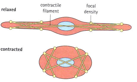
Unlike skeletal muscles they are 2 to 10 µm and have only one nuclei. They contain similar components to both cardiac and skeletal muscle cells; myosin, actin and tropomyosin but they do not have troponin. Instead, the myosin-head binding sites on the actin filaments are blocked by the protein calmodulin. When calcium ions are released from the extracellular fluid, 4 calcium ions bind to the protein calmodulin. This activates an enzyme - myosin light chain kinase - which phosphorylates the regulatory light chain myosin-heads. This activates myosin ATPase activity enabling cross-bridge formation and consequently muscular contraction.[7] The non-striated cells contain more actin than myosin in the fibre composition. Therefore, there is a larger proportion of thin filaments than thick filaments in smooth muscles than striated muscles. The actin and myosin are arranged in a diagonal web like structure around the call and are attatched to the cell membrane via dense bodies and protein attatchment plaques. The contractile fobres are in les organised bundles rather than the sacromeres observer in skeletal muscles. The resulting contraction moves the cell in varios directions.Smoth muscle is often layered in many different directions. [8]
The mode of control is mostly governed by the autonomic nervous system, meaning it is an involuntary control. Whereas, the skeletal muscles are innervated by the somatic nervous system control. The neuron can make contact with the smooth muscle cell at many points on the cell. This forms a swelling called a varicosity which contains the components for vesicular neurotransmitter release. The multiunit smooth muscle's cells each receive a nervous input and act independently to each other. The single unit muscle cells recieve a nervous input together and due to the many gap junctions electrical communication and take place. This allows the cells to act in unison[9]. Smooth muscle also respond to hormones and paracrines and consequently has to modulate multiple signals simultaneously, this results in differing electical behaviors. The variety of responce from these stimuli makes the muscle hard to work with.
Smooth muscles are able to undergo both muscle hypertrophy as well as muscle hyperplasia in response to increasing demands of heavier workloads. Hyperplasia is usually the major response. Muscle atrophy also occur in smooth muscles, as in the uterine smooth muscle after menopause, indicating that the status of uterine smooth muscle is under hormonal control [10].
References
- ↑ Raven, P.H. and Johnson, G.B. (1999) Biology(5th ed.) P915 WCB/McGraw-Hill
- ↑ Moffet, Moffett, Schauf(1993)Human Physiology, 2nd Edition, St. Louis, p313-314
- ↑ Stevens A. et al. (2005), Human Histology, Third Edition, Philadelphia, Elsevier Limited
- ↑ http://books.google.se/books?id=iOEQWGfiurYC&amp;amp;amp;amp;amp;amp;amp;amp;amp;amp;amp;amp;amp;amp;pg=PA175&amp;amp;amp;amp;amp;amp;amp;amp;amp;amp;amp;amp;amp;amp;lpg=PA174#v=onepage&amp;amp;amp;amp;amp;amp;amp;amp;amp;amp;amp;amp;amp;amp;q&amp;amp;amp;amp;amp;amp;amp;amp;amp;amp;amp;amp;amp;amp;f=false
- ↑ silverthorn 2010 Human phisiology, 5th edition pearson international chapter 12
- ↑ Bruce M. Koeppen and Bruce A Stanton (2008) Berne and Levy Physiology, 6th edition, Philadelphia: Moseby Elsevier.
- ↑ Guyton A, Hall J, 1997, Human Physiology and Mechanisms of Disease, 6th Edition, W.B. Saunders Company.
- ↑ silverthorn 2010, human phisiology, 5th edition pearson international
- ↑ Animal Physiology, Second Edition, Richard W.Hill Michigan State University, Gordon A. Wyse University of Massachusetts Amherst, Margaret Anderson Smith College,
- ↑ Stevens A. et al. (2005), Human Histology, Third Edition, Philadelphia, Elsevier Limited