DNA: Difference between revisions
No edit summary |
mNo edit summary |
||
| Line 9: | Line 9: | ||
The 2` deoxyribose sugar/phosphate group region is regarded as the 'backbone' of DNA strands due to its structural purpose, and the sequence of bases carries the gentic information. In order to produce a double stranded DNA structure, interactions occur between complementary bases. The complementary base pairs in DNA interact with one another via [[Hydrogen bonds|hydrogen bonds]]: A-T interactions consist of 2 intermolecular [[Hydrogen bonds|hydrogen bonds]], whereas G-C interactions consist of 3 intermolecular [[Hydrogen bonds|hydrogen bonds]]. These interactions form bridges between two DNA chains, thus creating a double stranded 'ladder' shaped structure. Each strand acts as a template for the other one in DNA replication. DNA is copied into [[MRNA|mRNA]] (messenger RNA) which carries the information from the original DNA template strand to be involved in protein synthesis. | The 2` deoxyribose sugar/phosphate group region is regarded as the 'backbone' of DNA strands due to its structural purpose, and the sequence of bases carries the gentic information. In order to produce a double stranded DNA structure, interactions occur between complementary bases. The complementary base pairs in DNA interact with one another via [[Hydrogen bonds|hydrogen bonds]]: A-T interactions consist of 2 intermolecular [[Hydrogen bonds|hydrogen bonds]], whereas G-C interactions consist of 3 intermolecular [[Hydrogen bonds|hydrogen bonds]]. These interactions form bridges between two DNA chains, thus creating a double stranded 'ladder' shaped structure. Each strand acts as a template for the other one in DNA replication. DNA is copied into [[MRNA|mRNA]] (messenger RNA) which carries the information from the original DNA template strand to be involved in protein synthesis. | ||
Despite many other theories, in 1953 James Watson and Frances Crick discovered the true structure of a double stranded DNA molecule to be a 'Double Helix'. This was solved as a result of 'stick-and-ball' models they created, along with utilising the work of fellow scientists [[Rosalind Franklin|Rosalind Franklin]] and [[Maurice Wilkins|Maurice Wilkins]] on [[X-ray crystallography|X-ray crystallography]]<ref>http://nobelprize.org/educational/medicine/dna_double_helix/readmore.html</ref> . The [[X-ray diffraction|X-ray diffraction]] photographs obtained from [[DNA|DNA]] fibres, displayed a unique X-shape, which illustrates a helical stucture, although they indicated a repeating structure of 3.4 Å apart per turn of the helix, each base is roated 36 degrees from the next one. The diameter of the helix is 20Â. They found that the sugar-phosphate backbone was on the outside and the bases are positioned on the inside of the helix <ref>http://www.chm.bris.ac.uk/motm/dna/dna.htm</ref><ref>J.Berg, J.Tymoczko, L.Stryer;, 113-115, 2012 Freeman; Biochemistry</ref>.<br> | Despite many other theories, in 1953 [[James_watson|James Watson]] and Frances Crick discovered the true structure of a double stranded DNA molecule to be a 'Double Helix'. This was solved as a result of 'stick-and-ball' models they created, along with utilising the work of fellow scientists [[Rosalind Franklin|Rosalind Franklin]] and [[Maurice Wilkins|Maurice Wilkins]] on [[X-ray crystallography|X-ray crystallography]]<ref>http://nobelprize.org/educational/medicine/dna_double_helix/readmore.html</ref> . The [[X-ray diffraction|X-ray diffraction]] photographs obtained from [[DNA|DNA]] fibres, displayed a unique X-shape, which illustrates a helical stucture, although they indicated a repeating structure of 3.4 Å apart per turn of the helix, each base is roated 36 degrees from the next one. The diameter of the helix is 20Â. They found that the sugar-phosphate backbone was on the outside and the bases are positioned on the inside of the helix <ref>http://www.chm.bris.ac.uk/motm/dna/dna.htm</ref><ref>J.Berg, J.Tymoczko, L.Stryer;, 113-115, 2012 Freeman; Biochemistry</ref>.<br> | ||
=== References === | === References === | ||
<references /><br> | <references /><br> | ||
Revision as of 15:12, 15 October 2012

DNA (deoxyribonucleic acid) is the genetic information found in the nuclei of most organisms. It is arranged into structures called chromosomes. The structure of DNA was identified as being a 'double-helix' by Watson and Crick in 1953. DNA is composed of 4 bases; the purines: adenine (A) and guanine (G) and the pyrimidines: thymine (T) and cytosine (C) [1]. These form complementary base pairs of AT and GC. DNA also contains a phosphate group connected to a deoxyribose sugar.
Structure of DNA
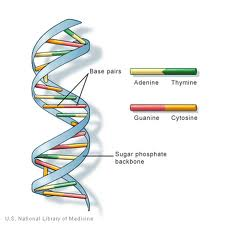
DNA strands are primarily composed of three repeating units: a 2` deoxyribose sugar (A five (pentose) carbon sugar similar to that of ribose sugar found in RNA. Its chemical formula is C5H10O4), a phosphate group (contains one phosphorus atom, bonded to 4 oxygens and forms a phosphodiester bond, which connects 2 deoxyribose sugars together resulting in the formation of a chain) and a base (one from A, C, G or T, which forms a side chain branching from the 2` deoxyribose sugar).
The 2` deoxyribose sugar/phosphate group region is regarded as the 'backbone' of DNA strands due to its structural purpose, and the sequence of bases carries the gentic information. In order to produce a double stranded DNA structure, interactions occur between complementary bases. The complementary base pairs in DNA interact with one another via hydrogen bonds: A-T interactions consist of 2 intermolecular hydrogen bonds, whereas G-C interactions consist of 3 intermolecular hydrogen bonds. These interactions form bridges between two DNA chains, thus creating a double stranded 'ladder' shaped structure. Each strand acts as a template for the other one in DNA replication. DNA is copied into mRNA (messenger RNA) which carries the information from the original DNA template strand to be involved in protein synthesis.
Despite many other theories, in 1953 James Watson and Frances Crick discovered the true structure of a double stranded DNA molecule to be a 'Double Helix'. This was solved as a result of 'stick-and-ball' models they created, along with utilising the work of fellow scientists Rosalind Franklin and Maurice Wilkins on X-ray crystallography[2] . The X-ray diffraction photographs obtained from DNA fibres, displayed a unique X-shape, which illustrates a helical stucture, although they indicated a repeating structure of 3.4 Å apart per turn of the helix, each base is roated 36 degrees from the next one. The diameter of the helix is 20Â. They found that the sugar-phosphate backbone was on the outside and the bases are positioned on the inside of the helix [3][4].
References
- ↑ HARTL AND JONES,2009:41, GENETICS : ANALYSIS OF GENES AND GENOMES SEVENTH EDITION.
- ↑ http://nobelprize.org/educational/medicine/dna_double_helix/readmore.html
- ↑ http://www.chm.bris.ac.uk/motm/dna/dna.htm
- ↑ J.Berg, J.Tymoczko, L.Stryer;, 113-115, 2012 Freeman; Biochemistry