Glycogenolysis: Difference between revisions
mNo edit summary |
mNo edit summary |
||
| (23 intermediate revisions by 4 users not shown) | |||
| Line 1: | Line 1: | ||
== Definition | == Definition == | ||
Glycogenolysis is defined as | Glycogenolysis is defined as metabolism of [[Glycogen|glycogen]] polymers occured during fasting. The glycogen is broken down in the [[Liver|liver]], [[Kidney|kidney]] or [[Muscle|muscles]] into [[Glucose|glucose]] or to [[Glucose-6-phosphate|glucose-6-phosphate]] for use in [[Glycolysis|glycolysis]] pathway.<ref name="[1]">Silverthorn, D.U. (2013) Human Physiology, An Integrated Approach. Sixth Edition. United States of America: Pearson Education.</ref> | ||
== | == Steps of glycogenolysis (glycogen breakdown) == | ||
==== 1. Phosphorolysis/Shoterning of chains ==== | |||
Glycogen is a branched polymer of glucose | Glycogen is a branched polymer of glucose units in chains are linked by [[1,4 glycosidic bonds|α-1,4-glycosidic bonds]] with a branch point created by [[1-6 glycosidic bond|α-1,6-glycosidic bonds]] at approximately every 10 residues of glucose. The key enzyme for glycogenolysis, [[Glycogen phosphorylase|glycogen phosphorylase]], will cleave the α-1,4-glycosidic bonds of the terminal glucose residues at the non-reducing end of glycogen (i.e. the end of the glycogen molecule with a free 4-OH group (''refer to Figure 1'') until only four glucosyl units remain on each chain before a branch point. | ||
'''[[Image:Glycogen structure.png]]''' | '''[[Image:Glycogen structure.png|Note the nonreducing end of the glycogen (the end with a free 4-OH group)]]''' | ||
'' | ''Figure 1 Structure of Glycogen'''''<ref name="[2]">Berg, J.M., Tymoczko, J.L and Stryer, L. (2012) Biochemistry. Seventh Edition. New York: W.H . Freeman and Company</ref>''' | ||
[[ | [[Orthophosphate|Orthophosphate]] (P<sub>i</sub>), an [[Inorganic phosphate|inorganic phosphate]], cleaves the the glycosidic bond between C1 of the terminal residue and C4 of the adjacent glycogen molecule via phosphorolysis to yield [[Glucose 1-phosphate|glucose 1-phosphate]]. The α configuration at C1 is retained even after the glycogen phosphorylase cleaves the bond between the C1 carbon atom and the glycosidic oxygen atom ''(refer to Figure 3).'' | ||
[[Image:Equation-Glycogen(n residues) to n-1 residues.png|Glycogen is attacked by an inorganic phosphate molecule (Pi)]] | |||
''' | ''Figure 2 Equation for phosphorolysis'''''<ref name="[2]">Berg, J.M., Tymoczko, J.L and Stryer, L. (2012) Biochemistry. Seventh Edition. New York: W.H . Freeman and Company</ref>''' | ||
[[Image:Glycogen.png|A molecule of glucose 6-phosphate is being produced in phosphorolysis.]] | |||
Glycogen phosphorylase can only carry out the glycogen breakdown process by itself until a limited extent before encountering an obstacle | ''Figure 3 Phosphorolysis by an orthophosphate (inorganic phosphate)'''''<ref name="[2]">Berg, J.M., Tymoczko, J.L and Stryer, L. (2012) Biochemistry. Seventh Edition. New York: W.H . Freeman and Company</ref>''' | ||
==== 2. Debranching/Removal of branches ==== | |||
Glycogen phosphorylase can only carry out the glycogen breakdown process by itself until a limited extent before encountering an obstacle. <ref name="[2]">Berg, J.M., Tymoczko, J.L and Stryer, L. (2012) Biochemistry. Seventh Edition. New York: W.H . Freeman and Company</ref> When phophorylase reaches a terminal residue four residues away from a branch point (i.e. after release of six glucose molecules), it will stop cleaving and the α-1,6 linkages are not susceptible to cleavage by phosphorylase. The branches of the glycogen molecule are removed by the debranching enzyme, a single bifunctional protein with two enzymic activities. The debranching enzyme can act as a [[Transferases|transferase]] as well as an [[Α-1,6-glucosidase|α-1,6-glucosidase]] to aid the continued degradation by phosphorylase. A block of three glycosyl residues from one outer branch was shifted by the transferase. The remaining single glucose molecule has a α-1,6-glycosidic bond joined to the glycogen molecule. The debranching enzyme, which act as α-1,6-glucosidase will cleave the linkage and results in the release of a free glucose molecule. The glycolytic enzyme, [[Hexokinase|hexokinase]] will phosphorylate this free glucose molecule. Thus, the net result is a linear structure which can be continue degraded by glycogen phosphorylase. | |||
[[Image:Glycogen remodelling.png]] | [[Image:Glycogen remodelling.png]] | ||
''Figure 4 Glycogen remodelling in glycogenolysis (Step 1 and step 2)'''''<ref name="[2]">Berg, J.M., Tymoczko, J.L and Stryer, L. (2012) Biochemistry. Seventh Edition. New York: W.H . Freeman and Company</ref>''' | |||
==== 3. Recovery ==== | |||
As in the glycolysis pathway, phosphoglucomutase is used to convert glucose 1-phosphate formed in the cleavage of glycogen into glucose 6-phosphate to enter the metabolic mainstream. | As in the [[Glycolysis|glycolysis]] pathway, [[Phosphoglucomutase|phosphoglucomutase]] is used to convert glucose 1-phosphate formed in the cleavage of glycogen into glucose 6-phosphate to enter the metabolic mainstream. | ||
'' | ==== 4''. ''Release ==== | ||
This process will only occur in liver. In contrast with glucose, the phosphorylated glucose produce in the glycogen breakdown is not readily to be transported out of the cell. The liver contains a hydrolytic enzyme, glucose 6-phosphatase, located on the luminal side of smooth endoplasmic reticulum, which convert the glucose 6-phosphate into glucose by cleaving the phosphoryl group. | This process will only occur in liver. In contrast with glucose, the phosphorylated glucose produce in the glycogen breakdown is not readily to be transported out of the cell. The liver contains a hydrolytic enzyme, glucose 6-phosphatase, located on the luminal side of smooth endoplasmic reticulum, which convert the glucose 6-phosphate into glucose by cleaving the phosphoryl group. | ||
Muscle cells, lack the enzyme that can make glucose from glucose 6-phosphate. Skeletal muscle, in fasted state, will metabolise glucose 6-phosphate to either pyruvate (aerobic conditions) or lactate (anaerobic conditions). | Muscle cells, lack the enzyme that can make glucose from glucose 6-phosphate. Skeletal muscle, in fasted state, will metabolise glucose 6-phosphate to either [[Pyruvate|pyruvate]] ([[Aerobic|aerobic]] conditions) or [[Lactate|lactate]] ([[Anaerobic|anaerobic]] conditions). '''<ref name="[1]">Silverthorn, D.U. (2013) Human Physiology, An Integrated Approach. Sixth Edition. United States of America: Pearson Education.</ref>''' | ||
== Effect of hormone on glycogenolysis == | |||
Metabolic effects of [[Glucagon|glucagon]] on glycogen breakdown – Primary target of glucagon is liver as glucagon receptors are not found on skeletal muscle. <ref name="[3]">Ferrier, D.R. (2011) Lippincott’s Illustrated Reviews: Biochemistry. Sixth Edition. Philadelphia: Lippincott Williams and Wilkins.</ref> In fasted state, glucagon will be produced in order to increase the plasma concentration of glucose. Under the influence of glucagon, enzymes that break down glycogen become more active but the enzymes for glycogen synthesis ([[Glycogenesis|glycogenesis]]) will become less active or inhibited. | |||
< | Stimulation of glycogen breakdown by [[Epinephrine|epinephrine]]/adrenaline – An epinephrine/adrenaline molecule binds to a β-adrenergic receptor on the plasma membrane of a liver or muscle cell. The [[G-protein|G protein]] neighbouring the receptor is activated and it in turn activate stimulate the [[Adenylyl cyclase|adenylyl cyclase]]. Adenylyl cyclase will then generate [[CAMP|cAMP]] from [[ATP|ATP]]. The increase of cAMP in the [[Cytosol|cytosol]] will activates the [[Protein kinase A|protein kinase A ]](PKA). PKA then increases the [[Phosphorylation|phosphorylation]] of the enzyme phosphorylase kinase. Glycogen phosphorylase will then be converted from phosphorylase b, the less active form, to phosphorylase a, the more active form. The rate of glycogen breakdown will increase significantly. <ref name="[4]">Hardin, J., Bertoni, G. and Kleinsmith, L.J. (2012) Becker’s World of the Cell. Eighth Edition. San Francisco: Pearson Benjamin Cummings.</ref> | ||
== References == | |||
<references /> | |||
Latest revision as of 00:58, 20 October 2015
Definition
Glycogenolysis is defined as metabolism of glycogen polymers occured during fasting. The glycogen is broken down in the liver, kidney or muscles into glucose or to glucose-6-phosphate for use in glycolysis pathway.[1]
Steps of glycogenolysis (glycogen breakdown)
1. Phosphorolysis/Shoterning of chains
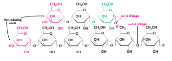
Glycogen is a branched polymer of glucose units in chains are linked by α-1,4-glycosidic bonds with a branch point created by α-1,6-glycosidic bonds at approximately every 10 residues of glucose. The key enzyme for glycogenolysis, glycogen phosphorylase, will cleave the α-1,4-glycosidic bonds of the terminal glucose residues at the non-reducing end of glycogen (i.e. the end of the glycogen molecule with a free 4-OH group (refer to Figure 1) until only four glucosyl units remain on each chain before a branch point.
Figure 1 Structure of Glycogen[2]
Orthophosphate (Pi), an inorganic phosphate, cleaves the the glycosidic bond between C1 of the terminal residue and C4 of the adjacent glycogen molecule via phosphorolysis to yield glucose 1-phosphate. The α configuration at C1 is retained even after the glycogen phosphorylase cleaves the bond between the C1 carbon atom and the glycosidic oxygen atom (refer to Figure 3).
Figure 2 Equation for phosphorolysis[2]
Figure 3 Phosphorolysis by an orthophosphate (inorganic phosphate)[2]
2. Debranching/Removal of branches
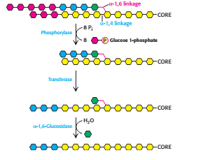
Glycogen phosphorylase can only carry out the glycogen breakdown process by itself until a limited extent before encountering an obstacle. [2] When phophorylase reaches a terminal residue four residues away from a branch point (i.e. after release of six glucose molecules), it will stop cleaving and the α-1,6 linkages are not susceptible to cleavage by phosphorylase. The branches of the glycogen molecule are removed by the debranching enzyme, a single bifunctional protein with two enzymic activities. The debranching enzyme can act as a transferase as well as an α-1,6-glucosidase to aid the continued degradation by phosphorylase. A block of three glycosyl residues from one outer branch was shifted by the transferase. The remaining single glucose molecule has a α-1,6-glycosidic bond joined to the glycogen molecule. The debranching enzyme, which act as α-1,6-glucosidase will cleave the linkage and results in the release of a free glucose molecule. The glycolytic enzyme, hexokinase will phosphorylate this free glucose molecule. Thus, the net result is a linear structure which can be continue degraded by glycogen phosphorylase.
Figure 4 Glycogen remodelling in glycogenolysis (Step 1 and step 2)[2]
3. Recovery
As in the glycolysis pathway, phosphoglucomutase is used to convert glucose 1-phosphate formed in the cleavage of glycogen into glucose 6-phosphate to enter the metabolic mainstream.
4. Release
This process will only occur in liver. In contrast with glucose, the phosphorylated glucose produce in the glycogen breakdown is not readily to be transported out of the cell. The liver contains a hydrolytic enzyme, glucose 6-phosphatase, located on the luminal side of smooth endoplasmic reticulum, which convert the glucose 6-phosphate into glucose by cleaving the phosphoryl group.
Muscle cells, lack the enzyme that can make glucose from glucose 6-phosphate. Skeletal muscle, in fasted state, will metabolise glucose 6-phosphate to either pyruvate (aerobic conditions) or lactate (anaerobic conditions). [1]
Effect of hormone on glycogenolysis
Metabolic effects of glucagon on glycogen breakdown – Primary target of glucagon is liver as glucagon receptors are not found on skeletal muscle. [3] In fasted state, glucagon will be produced in order to increase the plasma concentration of glucose. Under the influence of glucagon, enzymes that break down glycogen become more active but the enzymes for glycogen synthesis (glycogenesis) will become less active or inhibited.
Stimulation of glycogen breakdown by epinephrine/adrenaline – An epinephrine/adrenaline molecule binds to a β-adrenergic receptor on the plasma membrane of a liver or muscle cell. The G protein neighbouring the receptor is activated and it in turn activate stimulate the adenylyl cyclase. Adenylyl cyclase will then generate cAMP from ATP. The increase of cAMP in the cytosol will activates the protein kinase A (PKA). PKA then increases the phosphorylation of the enzyme phosphorylase kinase. Glycogen phosphorylase will then be converted from phosphorylase b, the less active form, to phosphorylase a, the more active form. The rate of glycogen breakdown will increase significantly. [4]
References
- ↑ 1.0 1.1 Silverthorn, D.U. (2013) Human Physiology, An Integrated Approach. Sixth Edition. United States of America: Pearson Education.
- ↑ 2.0 2.1 2.2 2.3 2.4 Berg, J.M., Tymoczko, J.L and Stryer, L. (2012) Biochemistry. Seventh Edition. New York: W.H . Freeman and Company
- ↑ Ferrier, D.R. (2011) Lippincott’s Illustrated Reviews: Biochemistry. Sixth Edition. Philadelphia: Lippincott Williams and Wilkins.
- ↑ Hardin, J., Bertoni, G. and Kleinsmith, L.J. (2012) Becker’s World of the Cell. Eighth Edition. San Francisco: Pearson Benjamin Cummings.


